. 2004 Oct 12;101(41):14871-6.
doi: 10.1073/pnas.0406073101. Epub 2004 Oct 4.

Distinct endothelial phenotypes evoked by arterial waveforms derived from atherosclerosis-susceptible and -resistant regions of human vasculature

Affiliations

Distinct endothelial phenotypes evoked by arterial waveforms derived from atherosclerosis-susceptible and -resistant regions of human vasculature

Guohao Dai et al. Proc Natl Acad Sci U S A. .

Abstract

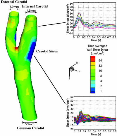
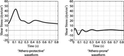
Atherosclerotic lesion localization to regions of disturbed flow within certain arterial geometries, in humans and experimental animals, suggests an important role for local hemodynamic forces in atherogenesis. To explore how endothelial cells (EC) acquire functional/dysfunctional phenotypes in response to vascular region-specific flow patterns, we have used an in vitro dynamic flow system to accurately reproduce arterial shear stress waveforms on cultured human EC and have examined the effects on EC gene expression by using a high-throughput transcriptional profiling approach. The flow patterns in the carotid artery bifurcations of several normal human subjects were characterized by using 3D flow analysis based on actual vascular geometries and blood flow profiles. Two prototypic arterial waveforms, "athero-prone" and "athero-protective," were defined as representative of the wall shear stresses in two distinct regions of the carotid artery (carotid sinus and distal internal carotid artery) that are typically "susceptible" or "resistant," respectively, to atherosclerotic lesion development. These two waveforms were applied to cultured EC, and cDNA microarrays were used to analyze the differential patterns of EC gene expression. In addition, the differential effects of athero-prone vs. athero-protective waveforms were further characterized on several parameters of EC structure and function, including actin cytoskeletal organization, expression and localization of junctional proteins, activation of the NF-kappaB transcriptional pathway, and expression of proinflammatory cytokines and adhesion molecules. These global gene expression patterns and functional data reveal a distinct phenotypic modulation in response to the wall shear stresses present in atherosclerosis-susceptible vs. atherosclerosis-resistant human arterial geometries.


Figures

Fig. 1.
Fig. 1.
Hemodynamics in human carotid bifurcation manifests distinct site specificity. The left carotid anatomy of a normal human subject (27-year-old male) was reconstructed from noninvasive MRI measurements. The color-coded map shown in this carotid model displays the time-averaged shear stress magnitude at different points along the vascular wall. Regions in the distal internal carotid artery and carotid sinus were selected, and the time-dependent shear stress values along the mean flow direction from multiple individual points within each region were plotted for one cardiac cycle. For the carotid sinus, all of the finite element surface grid points within a 4-mm diameter circle in the sinus region (total 199 grid points) were chosen, and the time-dependent shear stresses along the mean flow direction from individual points were plotted (Lower Right). Each waveform depicted thus represents the time-dependent wall shear stress profile at a given point in each region of interest.
Fig. 2.
Fig. 2.
Selection of prototypic athero-prone (Right) and athero-protective (Left) waveforms. Two waveforms were chosen as representative of the hemodynamics corresponding to an atherosclerosis-susceptible and an atherosclerosis-resistant region of the human carotid artery. The waveform with the highest OSI (0.45; corresponding to point 14 in Fig. 6) was chosen from the carotid sinus to represent the wall shear stress fluctuations in this atherosclerosis-susceptible region. Another shear stress waveform from the distal internal carotid artery was chosen as representative of the wall shear stress profile in an atherosclerosis-resistant region. These two waveforms (designated athero-prone and athero-protective, respectively) were then applied to cultured human EC by using the dynamic flow system as described in Materials and Methods.
Fig. 3.
Fig. 3.
Actin cytoskeletal organization and subcellular localization of Cx37 and Cx43 are regulated differentially by shear stress waveforms. EC monolayers were immunofluorescently stained for F-actin, Cx37, and Cx43, after 24 h of exposure to static (no flow) and athero-prone and athero-protective waveforms. (Top) Nuclei were stained with SYTOX (Molecular Probes) (red) (×40).
Fig. 4.
Fig. 4.
Athero-prone waveform stimulation results in enhanced IL-8 production and NF-κB nuclear translocation. (a) IL-8 mRNA expression was measured by TaqMan RT-PCR (n = 3) at 24 h. All data are normalized to the static (no flow) condition. (b) Endothelial IL-8 secretion was measured by ELISA (n = 3). EC were cultured under static (no flow) or athero-prone and athero-protective waveform for 24 h. The supernant was collected from the outflow port of the dynamic flow device every 2 h, and IL-8 protein was measured by ELISA (see Materials and Methods). (c) Immunofluorescent staining of NF-κB (p65) (×40). EC were stained with antibodies to p65 after 24 h exposure to static (no flow) and athero-prone and athero-protective waveform. (d) Western blot analysis of NF-κB (p65) protein levels. HUVEC were cultured under static, athero-prone, or athero-protective flow conditions for 24 h, then treated with IL-1β (1 units/ml) for 1 h under static condition. Cytoplasmic and nuclear protein fractions were isolated and p65 protein was analyzed by Western blotting. The blot was then stripped and reprobed for α-tubulin and histone H3. Lanes 1, static (no flow); lanes 2, athero-prone; lanes 3, athero-protective condition.
Fig. 5.
Fig. 5.
Preconditioning with an athero-protective waveform suppresses IL-1β-inducible VCAM-1 expression. (a and b) Flow cytometry analysis of EC surface expression of VCAM-1 (a) and E-selectin (b). HUVEC were preconditioned by exposure to static (no flow), athero-prone, or athero-protective conditions for 24 h (a Upper and b Upper), and then challenged with IL-1β (1 units/ml) for 6 h (for VCAM-1, a Lower) or 4 h (for E-selectin, b Lower) under static conditions, followed by flow cytometric analysis of surface VCAM-1 or E-selectin expression. (c) Western blot analysis of VCAM-1 and E-selectin protein expression. HUVEC preconditioned by exposure to static (lanes 1; no flow) or athero-prone (lanes 2) and athero-protective (lanes 3) conditions for 24 h were challenged with IL-1β (1 units/ml) for 4 h under static condition. Cells were lysed, and VCAM-1 and E-selectin proteins were analyzed by Western blotting.

Comment in

References

    1. Ross, R. (1999) N. Engl. J. Med. 340, 115-126. - PubMed
    1. VanderLaan, P. A., Reardon, C. A. & Getz, G. S. (2004) Arterioscler. Thromb. Vasc. Biol. 24, 12-22. - PubMed
    1. Wissler, R. W. & Strong, J. P. (1998) Am. J. Pathol. 153, 1023-1033. - PMC - PubMed
    1. Corti, R., Fuster, V., Badimon, J. J., Hutter, R. & Fayad, Z. A. (2001) Ann. N.Y. Acad. Sci. 947, 181-195; discussion 195-198. - PubMed
    1. Ku, D. N., Giddens, D. P., Zarins, C. K. & Glagov, S. (1985) Arteriosclerosis 5, 293-302. - PubMed

Publication types

Substances

LinkOut - more resources