. 2004 Nov 26;279(48):50176-80.
doi: 10.1074/jbc.C400412200. Epub 2004 Oct 5.

GSK-3 phosphorylation of the Alzheimer epitope within collapsin response mediator proteins regulates axon elongation in primary neurons

Affiliations

GSK-3 phosphorylation of the Alzheimer epitope within collapsin response mediator proteins regulates axon elongation in primary neurons

Adam R Cole et al. J Biol Chem. .

Abstract

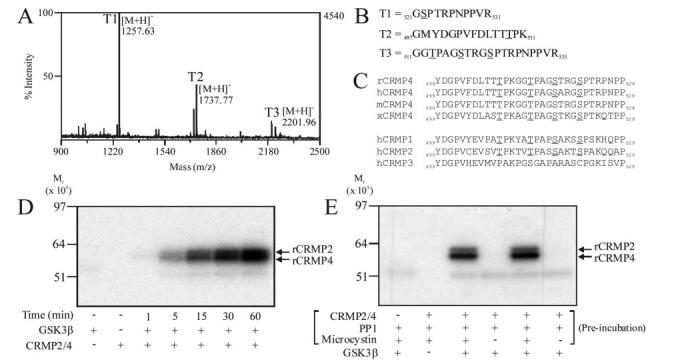
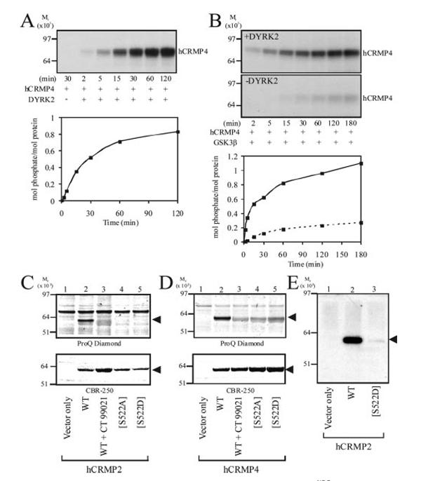
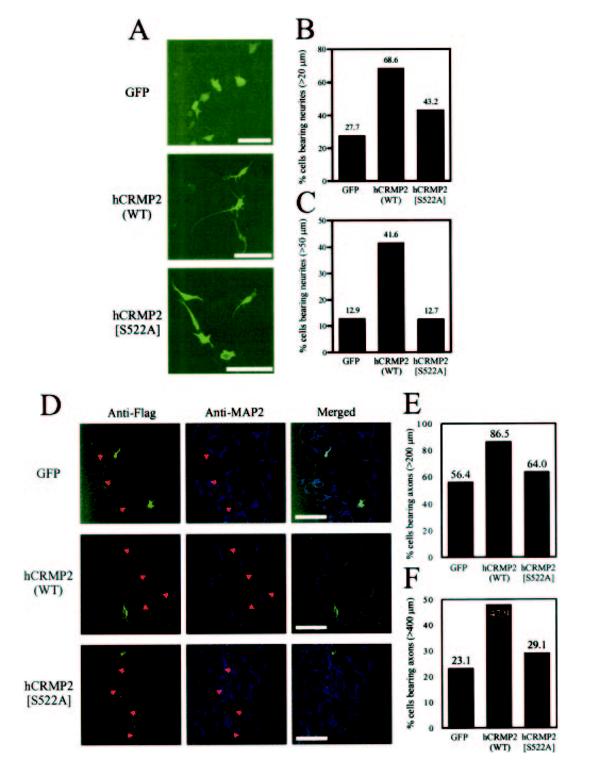
Elevated glycogen synthase kinase-3 (GSK-3) activity is associated with Alzheimer disease. We have found that collapsin response mediator proteins (CRMP) 2 and 4 are physiological substrates of GSK-3. The amino acids targeted by GSK-3 comprise a hyperphosphorylated epitope first identified in plaques isolated from Alzheimer brain. Expression of wild type CRMP2 in primary hippocampal neurons or SH-SY5Y neuroblastoma cells promotes axon elongation. However, a GSK-3-insensitive CRMP2 mutant has dramatically reduced ability to promote axon elongation, a similar effect to pharmacological inhibition of GSK-3. Hence, we propose that phosphorylation of CRMP proteins by GSK-3 regulates axon elongation. This work provides a direct connection between hyperphosphorylation of these residues and elevated GSK-3 activity, both of which are observed in Alzheimer brain.


Figures

FIG.1
FIG.1
Identification of the residues in rCRMP phosphorylated by GSK-3β. Purified rCRMP was phosphorylated using GSK-3β and then subjected to SDS-PAGE. Phosphorylated tryptic peptides were isolated using IMAC chromatography and identified using MALDI-TOF mass spectrometry. A, MS of tryptic phospho-peptides from rCRMP4. B, peptide identification of T1, T2, and T3. C, sequence alignment of residues Tyr499 to Pro529 of rCRMP4 with other species (rat, human, mouse, Xenopus), as well as other CRMP isoforms. Phosphorylated residues are underlined. D, purified rat brain CRMP2/4 was labeled for the times indicated using GSK-3β and the reactions subjected to SDS-PAGE, electrotransferred to nitrocellulose membrane, and autoradiographed. E, purified rCRMP2/4 was preincubated with the protein phosphatase PP1, with or without the phosphatase inhibitor microcystin as indicated. Microcystin was then added to the tubes that did not already contain it followed by 5 milliunits of GSK-3β and [γ-32P]ATP. Phosphate incorporated in each reaction is presented.
FIG.2
FIG.2
GST-hCRMP4 requires priming at Ser522 for subsequent phosphorylation by GSK-3β. A, GST-hCRMP4 was incubated with DYRK2 and [γ-32P]ATP for the times indicated, prior to SDS-PAGE and autoradiography. Stoichiometry of phosphorylation at each time point is presented below the autoradiograph. B, GST-hCRMP4 was incubated with (upper panel) or without (lower panel) DYRK2 and non-radioactive ATP for 30 min. DYRK2 was removed using Ni2+-agarose and the supernatant incubated with His6-GSK-3β and [γ-32P]ATP. Aliquots were removed and analyzed as described for A. The stiochiometry of phosphorylation is presented below the autoradiograph (dashed line in the absence of DYRK2 preincubation). C, HEK293 cells were transfected with expression vectors as indicated and then incubated with the GSK-3 inhibitor CT-99021 (lane 3) or Me2SO. FLAG-tagged hCRMP2 was immunoprecipitated, subjected to SDS-PAGE, and phosphorylated proteins detected using ProQ Diamond phospho-specific stain (upper panel). Gels were subsequently stained with CBR-250 to detect protein content (lower panel). D, HEK293 cells were transfected with the equivalent constructs for hCRMP4 and analyzed as described for C. E, HEK293 cells transfected with the indicated expression vectors were lysed and hCRMP2 proteins immunoprecipitated subjected to Western blotting using an antibody that specifically recognizes hCRMP2 only when phosphorylated at both Thr509 and Thr514. hCRMP proteins in C, D, and E are indicated by arrows.
FIG.3
FIG.3
Phosphorylation of hCRMP2 by GSK-3 is required for neurite formation and elongation. A, SH-SY5Y cells were transfected with expression vectors containing GFP, hCRMP2, or hCRMP2[S522A] and incubated at 37 °C for 60 h. Cells transfected with hCRMP2 or hCRMP2[S522A] were detected using an anti-FLAG monoclonal antibody, followed by treatment with an Alexa488-conjugated secondary antibody, and neurite lengths were subsequently analyzed using LSM 510 image analysis software. The scale bar is equivalent to 100 μm. Cells bearing neurites greater than 200 μm (B) or 50 μm (C)in length were determined as a percentage of the total number of cells analyzed for each sample group (n > 100 in each group). D, confocal images of hippocampal neurons transfected with expression vectors containing GFP, hCRMP2, or hCRMP2[S522A]. Cells transfected with hCRMP2 or hCRMP2[S522A] were detected using an anti-FLAG antibody and neurones identified with an antibody that recognizes the somato-dendritic marker protein, MAP2. Axon length was obtained using LSM 510 software. The scale bar is equivalent to 100 μm. The proportion of cells with axons greater than 200 μm (E) or 400 μm (F)in length is presented for each sample group. Average axon length is given in the text.

References

    1. Woodgett JR. EMBO J. 1990;9:2431–2438. - PMC - PubMed
    1. Frame S, Cohen P. Biochem J. 2001;359:1–16. - PMC - PubMed
    1. Doble BW, Woodgett JR. J Cell Sci. 2003;116:1175–1186. - PMC - PubMed
    1. Sutherland C, Leighton IA, Cohen P. Biochem J. 1993;296:15–19. - PMC - PubMed
    1. Cole A, Frame S, Cohen P. Biochem J. 2004;377:249–255. - PMC - PubMed

Publication types

Substances