. 2004 Oct;114(7):898-907.
doi: 10.1172/JCI21152.

Vasohibin as an endothelium-derived negative feedback regulator of angiogenesis

Affiliations

Vasohibin as an endothelium-derived negative feedback regulator of angiogenesis

Kazuhide Watanabe et al. J Clin Invest. 2004 Oct.

Abstract

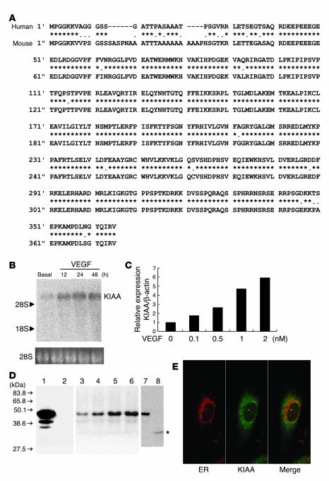
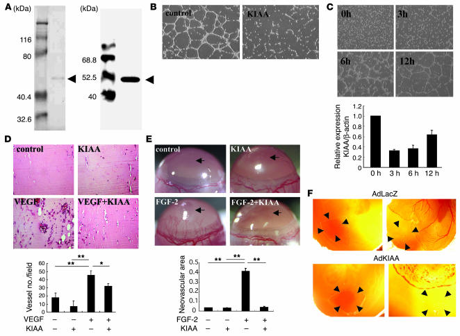
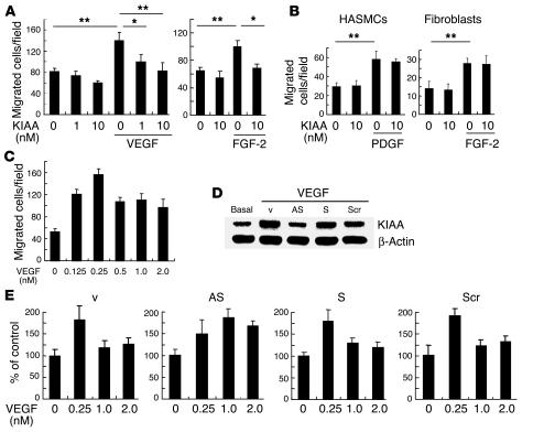
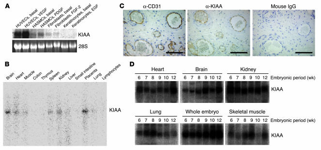
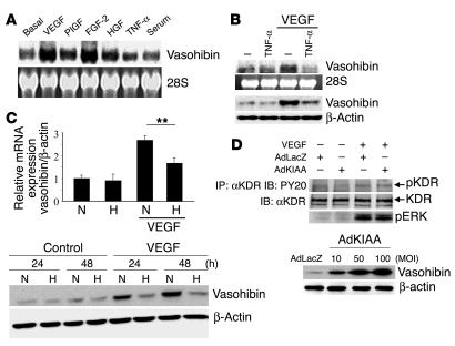
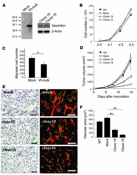
Negative feedback is a crucial physiological regulatory mechanism, but no such regulator of angiogenesis has been established. Here we report a novel angiogenesis inhibitor that is induced in endothelial cells (ECs) by angiogenic factors and inhibits angiogenesis in an autocrine manner. We have performed cDNA microarray analysis to survey VEGF-inducible genes in human ECs. We characterized one such gene, KIAA1036, whose function had been uncharacterized. The recombinant protein inhibited migration, proliferation, and network formation by ECs as well as angiogenesis in vivo. This inhibitory effect was selective to ECs, as the protein did not affect the migration of smooth muscle cells or fibroblasts. Specific elimination of the expression of KIAA1036 in ECs restored their responsiveness to a higher concentration of VEGF. The expression of KIAA1036 was selective to ECs, and hypoxia or TNF-alpha abrogated its inducible expression. As this molecule is preferentially expressed in ECs, we designated it "vasohibin." Transfection of Lewis lung carcinoma cells with the vasohibin gene did not affect the proliferation of cancer cells in vitro, but did inhibit tumor growth and tumor angiogenesis in vivo. We propose vasohibin to be an endothelium-derived negative feedback regulator of angiogenesis.


Figures

Figure 1
Figure 1
KIAA1036 is an endothelium-derived VEGF-inducible secretory protein. (A) The deduced amino acid sequences of the human and mouse KIAA1036 (KIAA) proteins are shown. Asterisks indicate identical amino acids between human and mouse. (B) A single KIAA1036 mRNA was induced by VEGF. HUVECs were stimulated with VEGF (1 nM) for the indicated periods and then Northern blotting was performed. (C) VEGF increased KIAA1036 mRNA in a concentration-dependent manner. HUVECs were stimulated with the indicated concentration of VEGF for 24 hours and then real-time RT-PCR was performed. (D) KIAA1036 protein was synthesized and secreted. GM7373 cells transfected with KIAA1036 gene were lysed. Equal amounts of protein were applied to lane 1 and lane 2, and transferred to the filter. The filter was then separated into 2 parts. Western blotting was performed with anti_KIAA1036 mAb (lane 1). Prior to Western blotting, anti_KIAA1036 mAb was absorbed with antigen peptide (lane 2). HUVECs were stimulated with VEGF (1 nM) for the following periods and were lysed for Western blotting: lane 3, 0 hours; lane 4, 12 hours; lane 5, 24 hours; lane 6, 48 hours. HUVECs were cultured for 3 days in the growth medium and then cells were lysed for Western blotting (lane 7). After this incubation, the medium was collected and concentrated. Five hundred microliters of concentrated medium was subjected to immunoprecipitation followed by Western blotting (lane 8). Asterisk indicates protein in the medium. (E) KIAA1036 protein does not colocalize with ER. HUVECs in the growth medium were used for the immunostaining of calnexin (red) and KIAA1036 protein (green).
Figure 2
Figure 2
KIAA1036 inhibits angiogenesis. (A) Preparation of KIAA1036 protein. SDS-PAGE/Coomassie brilliant blue staining is shown on the left and Western blotting on the right. Arrows indicate KIAA1036. (B) Effect of KIAA1036 on network formation by ECs. HUVECs were plated on Matrigel in the absence or presence of KIAA1036 (10 nM). (C) Downregulation of KIAA1036 during network formation on Matrigel. HUVECs were plated on Matrigel, and after the indicated period of incubation, total RNA was obtained and real-time RT-PCR was performed. Values are expressed as mean ± SD of 3 samples. (D) Matrigel implantation analysis was performed as described in Methods. The vessel number per low-power field in 3 different fields was counted for each sample. Values are expressed as mean ± SD of 5 animals. (E) Mouse corneal micropocket assay was performed as described in Methods. Arrows indicate the site where pellets were implanted. Neovascular area (mm2) was determined using NIH Image. Values are expressed as mean ± SD of 5 eyes. (F) CAM assay using adenovirus vectors was performed as described in Methods. The nylon mesh containing adenovirus/Matrigel mixture was placed on the peripheral zone of the CAM, where vascular structure would not appear (left panels). Four days after adenovirus infection, vascular formation was evaluated by macroscopic observation (right). Arrowheads indicate the site where the nylon mesh was placed. *P < 0.05, **P < 0.01.
Figure 3
Figure 3
KIAA1036 may act as a negative feedback regulator. (A) Effect of KIAA1036 on the migration of HUVECs. Migration of HUVECs was analyzed as described in Methods. The indicated concentrations of growth factors and/or KIAA1036 protein were placed in the lower chamber of the Transwell insert. Values are expressed as mean ± SD of 4 samples. (B) Effect of KIAA1036 on the migration of HASMCs or fibroblasts. Migration of HASMCs or fibroblasts was analyzed as described in Methods. The indicated combinations of PDGF, FGF-2, and KIAA1036 protein were placed in the lower chamber. Values are expressed as mean ± SD of 4 samples. (C) Bell-shaped pattern of the VEGF-stimulated migration of HUVECs. The indicated concentrations of VEGF were placed in the lower chamber and HUVECs were plated in the upper chamber. Values are expressed as mean ± SD of 4 samples. (D) Selective downregulation of KIAA1036 synthesis. HUVECs were incubated for 4 hours with 500 nM synthetic phosphorothioate ODNs or vehicle alone. Thereafter, HUVECs were stimulated with VEGF (1 nM) for 24 hours, and Western blotting for KIAA1036 was performed. AS, AS-ODN; S, S-ODN; Scr, Scr-ODN; v, vehicle. (E) Modulation of the bell-shaped pattern of the VEGF effect by KIAA1036 AS-ODNs. HUVECs were incubated for 4 hours with 500 nM phosphorothioate ODNs. Thereafter, HUVECs were subjected to the migration assay described above. Values are expressed as mean ± SD of 4 samples. *P < 0.05, **P < 0.01.
Figure 4
Figure 4
KIAA1036 is preferentially expressed in ECs. (A) Expression of KIAA1036 in cultured cells. Cells were preincubated in 0.1% FCS/α-MEM for 12 hours and then stimulated with growth factors as follows: HUVECs with VEGF (1 nM), HASMCs with PDGF (1 nM), human fibroblasts with FGF-2 (2 nM), and keratinocytes with EGF (1 nM). Thereafter, total RNA was obtained and Northern blotting for vasohibin was performed. (B) Expression of KIAA1036 in vivo was examined by multiple-tissue Northern blot. (C) Localization of KIAA1036 protein in the placenta. Sections of human placenta were subjected to immunostaining. Anti_human CD31 mAb, anti_KIAA1036 mAb, or mouse IgG was used as the primary Ab. Scale bars: 100 μm. (D) Expression of KIAA1036 in human embryo. Northern blotting for vasohibin was performed using a human developmental total RNA Northern blot.
Figure 5
Figure 5
Modulation of vasohibin expression and the effect of vasohibin on VEGF-stimulated signaling in HUVECs. (A) Induction of vasohibin. HUVECs were stimulated with VEGF (1 nM), PlGF (1 nM), FGF-2 (2 nM), HGF (1 nM), TNF-α (1 nM), or 10% serum for 24 hours. Thereafter, total RNA was obtained and Northern blotting for vasohibin was performed. (B) Effect of TNF-α on the induction of vasohibin by VEGF. HUVECs were stimulated with VEGF (1 nM) and/or TNF-α (1 nM). Thereafter, Northern blotting and Western blotting for vasohibin were performed. (C) Effect of hypoxia on the induction of vasohibin by VEGF. HUVECs were stimulated with VEGF (1 nM) under normoxic (N) or hypoxic (H) conditions. Upper panel: Total RNA was obtained and real-time RT-PCR of vasohibin was performed. Values are expressed as mean ± SD of 4 samples. **P < 0.01. Lower panel: Cell extract was obtained and Western blotting for vasohibin was performed. (D) Effect of vasohibin on VEGF-mediated KDR tyrosine phosphorylation or ERK1/2 activation of HUVECs. HUVECs were infected with AdLacZ or AdKIAA at an MOI of 100, and then stimulated with VEGF (10 ng/ml). VEGF-mediated KDR tyrosine phosphorylation or ERK1/2 activation was analyzed. Results shown in lower panel indicate that AdKIAA increased the synthesis of vasohibin in an MOI-dependent manner. IP, immunoprecipitation; IB, immunoblotting; pKDR, phosphorylated KDR.
Figure 6
Figure 6
Vasohibin suppresses tumor growth and tumor angiogenesis. (A) The synthesis of vasohibin protein in LLC cells. Cell extracts were prepared from mock or vasohibin transfectants (Vh-bulk) for Western blotting. Clone 16 and clone 19 were vasohibin-producing clones. (B) Effect of vasohibin on the proliferation of LLC cells in vitro. Proliferation of mock transfectants, vasohibin transfectants, clone 16, and clone 19 was determined. (C) Effect of secreted vasohibin from LLC cells on the migration of HUVECs. Mock or vasohibin transfectants were plated on the lower compartment of a modified Boyden chamber and the migration of HUVECs toward the lower chamber of the Transwell insert was analyzed. Values are expressed as mean ± SD of 4 samples. (D) Effect of vasohibin gene transfection on the growth of LLC cells in vivo. BDF1 mice were inoculated intradermally with LLC cells. Tumor volume was determined consecutively. (E) Effect of vasohibin gene transfection on tumor angiogenesis. Paraffin sections were prepared from tumors for the immunostaining of CD31; sections obtained on day 8 after inoculation are shown. Visualization with a DAKO LSAB+/HRP kit is shown at left, and that with streptavidin-Cy3 conjugate on the right. Yellow lines trace vascular lumens. Scale bars: 50 μm. (F) Quantitative analysis of tumor vascular area. Total vascular area per field was determined using NIH Image and compared. Values are expressed as mean ± SD of 6 random fields. *P < 0.05; **P < 0.01.

Comment in

References

    1. Folkman J. Angiogenesis in cancer, vascular, rheumatoid and other disease. Nat. Med. 1995;1:27–31. - PubMed
    1. Ribatti D, Vacca A, Presta M. The discovery of angiogenic factors: a historical review. Gen. Pharmacol. 2000;35:227–231. - PubMed
    1. Ferrara N. VEGF and the quest for tumour angiogenesis factors. Nat. Rev. Cancer. 2002;2:795–803. - PubMed
    1. Good DJ, et al. A tumor suppressor-dependent inhibitor of angiogenesis is immunologically and functionally indistinguishable from a fragment of thrombospondin. Proc. Natl. Acad. Sci. U. S. A. 1990;87:6624–6628. - PMC - PubMed
    1. Dameron KM, Volpert OV, Tainsky MA, Bouck NP. Control of angiogenesis in fibroblasts by p53 regulation of thrombospondin-1. Science. 1994;265:1582–1584. - PubMed

Publication types

MeSH terms