. 2004 Oct;114(7):944-52.
doi: 10.1172/JCI21786.

CEACAM1 modulates epidermal growth factor receptor--mediated cell proliferation

Affiliations

CEACAM1 modulates epidermal growth factor receptor--mediated cell proliferation

George A Abou-Rjaily et al. J Clin Invest. 2004 Oct.

Abstract

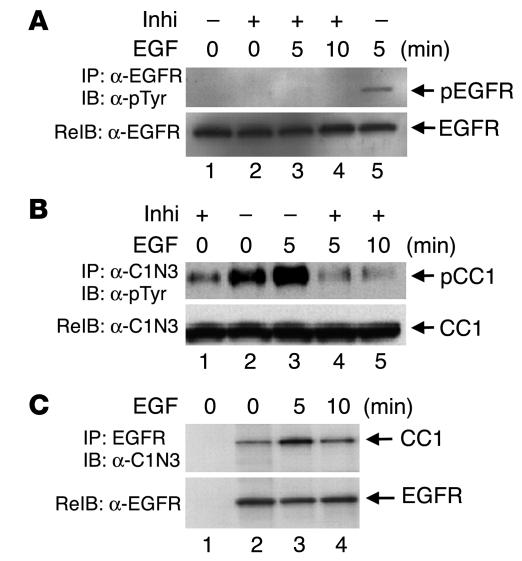
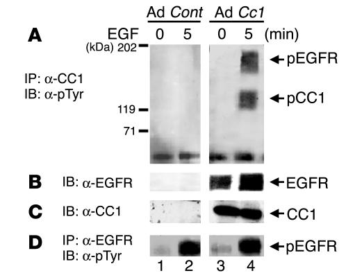
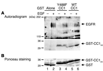
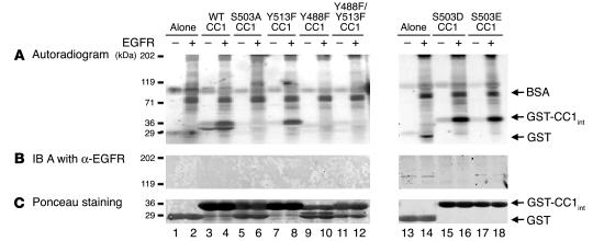
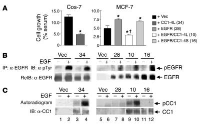
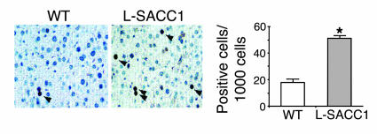
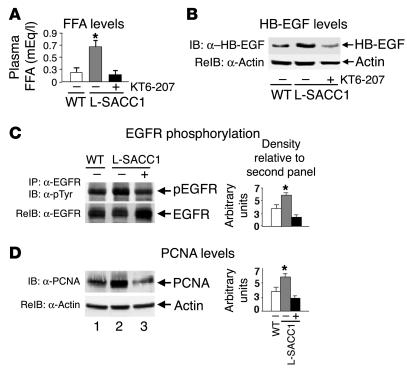
Phosphorylation of the cell adhesion protein CEACAM1 increases insulin sensitivity and decreases insulin-dependent mitogenesis in vivo. Here we show that CEACAM1 is a substrate of the EGFR and that upon being phosphorylated, CEACAM1 reduces EGFR-mediated growth of transfected Cos-7 and MCF-7 cells in response to EGF. Using transgenic mice overexpressing a phosphorylation-defective CEACAM1 mutant in liver (L-SACC1), we show that the effect of CEACAM1 on EGF-dependent cell proliferation is mediated by its ability to bind to and sequester Shc, thus uncoupling EGFR signaling from the ras/MAPK pathway. In L-SACC1 mice, we also show that impaired CEACAM1 phosphorylation leads to ligand-independent increase of EGFR-mediated cell proliferation. This appears to be secondary to visceral obesity and the metabolic syndrome, with increased levels of output of free fatty acids and heparin-binding EGF-like growth factor from the adipose tissue of the mice. Thus, L-SACC1 mice provide a model for the mechanistic link between increased cell proliferation in states of impaired metabolism and visceral obesity.


Figures

Figure 1
Figure 1
EGF induces CEACAM1-4L phosphorylation by EGFR in intact HT29p human colon cancer cells. Serum-starved HT29p cells were treated with buffer alone (_) or with the EGFR inhibitor PD168393 (Inhi; +) for 1 hour prior to treatment with EGF (100 nM) for 0_10 minutes. Equal amounts (100 μg) of cell lysates were immunoprecipitated (IP) with α-EGFR (A) or α-C1N3 (B) prior to analysis by SDS-PAGE and immunoblotting (IB) with α-pTyr for detection of tyrosine-phosphorylated proteins (pEGFR and pCC1) and re-immunoprobing (ReIB) with α-EGFR and α-C1N3 to account for the amount of these proteins in the immunoprecipitates. For examination of the CEACAM1-4L/EGFR association, 100 μg of cell lysates were immunoprecipitated with α-EGFR and immunoblotted with α-C1N3 (C, lanes 2_4). To account for nonspecific binding of proteins to agarose, equal amounts of proteins were incubated without the addition of antibody (C, lane 1). The results obtained were consistent in at least three experiments.
Figure 2
Figure 2
EGFR phosphorylates CEACAM1-4L in infected human MDA-MB468 breast cancer cells. MDA-MB468 breast cancer cells were infected with Ad Cc1 (lanes 3 and 4) or with Ad Cont (lanes 1 and 2) prior to EGF treatment. Equal amounts of proteins were immunoprecipitated with α-CC1 (A) or α-EGFR (D) prior to Western blot analysis with α-pTyr. (B) Proteins in A were reprobed with α-EGFR. (C) To account for the amount of CEACAM1-4L, amounts of proteins equal to those in A were applied on the same SDS-PAGE gel and were immunoblotted with α-CC1. The results obtained were consistent in at least 3 experiments.
Figure 3
Figure 3
CEACAM1-4L is a direct substrate of EGFR. (A) After treatment of MDA-MB468 cells with EGF (100 nM) (+) or buffer alone (_) for 5 minutes, cells were lysed and EGFR was extracted by immunoprecipitation with α-EGFR. GST fusion to intracellular peptides of wild type (WT CC1; lanes 5 and 6) or Y488F mutant (Y488F CC1; lanes 3 and 4) CEACAM1-4L (GST-CC1int) were incubated with equal amounts of EGFR immunoprecipitates in the presence of [γ-32P]ATP for 10 minutes prior to the addition of SDS buffer and analysis by 6_12% SDS-PAGE and autoradiography for the detection of phosphorylated proteins. Peptide-free GST was included as control to account for nonspecific association (Alone; lanes 1 and 2). (B) The amount of GST peptides from the experiment in A, measured by Ponceau S staining. The results obtained were consistent in at least three experiments. The two gels in A are from the same SDS-PAGE gel and differ in the exposure time of the autoradiogram for optimal visualization of both EGFR and GST bands.
Figure 4
Figure 4
CEACAM1-4L does not directly interact with EGFR. (A) Sepharose-coupled GST fusion to intracellular peptides of WT (lanes 3 and 4) or mutant (lanes 5_12 and 15_18) CEACAM1-4L were incubated with (+) or without (_) purified EGFR in the presence of [γ-32P]ATP. Peptide-free GST was included as control to account for nonspecific association (lanes 1, 2, 13, and 14). After protein binding, the Sepharose pellets were analyzed by 6_12% SDS-PAGE and autoradiography. (B) Proteins in A were immunoblotted with α-EGFR for examination of the association of EGFR with CEACAM1-4L. (C) The amount of GST peptides from the experiment in A was measured by Ponceau S staining. The results obtained were consistent in at least three experiments.
Figure 5
Figure 5
CEACAM1-4L downregulates cell growth in response to EGF. (A) Cos-7 and MCF-7 cells were stably transfected with vector alone (+ Vec) or with cDNA encoding rat CEACAM1-4L (+ CC1-4L), EGFR (+ EGFR), or both (+ EGFR/CC1-4L). Cells were also transfected with cDNA encoding EGFR and CEACAM1_4S (+ EGFR/CC1-4S). Numbers in parentheses (in key) denote the clone number. After being incubated for 24 hours in serum-containing complete medium (maximal growth) or in serum-free medium supplemented with 0.1% BSA either alone (basal growth) or with 100 nM EGF, cells were counted by the MTT method. EGF-induced cell growth was calculated as the percent maximal minus basal growth divided by the number of cells grown in complete medium. These experiments were performed in triplicate and were repeated at least three times per clone. Data represent the mean ± SD of repeated experiments. At least two stable clones were examined. *P < 0.05 versus Vec; P < 0.05 versus EGFR (clone 28). (B) For examination of EGFR phosphorylation, serum-starved cells were treated with EGF as described in Figure 1 and lysed and the EGFR immunoprecipitates were analyzed by SDS-PAGE, immunoblotting with α-pTyr for detection of tyrosine-phosphorylated EGFR, and re-immunoblotting with α-EGFR to account for the amount of EGFR in the immunoprecipitates. (C) For examination of CEACAM1-4L phosphorylation, cell lysates were lectin-purified prior to phosphorylation in the presence of EGF and [γ-32P]ATP, immunoprecipitation of CEACAM1-4L, and analysis of its phosphorylation by autoradiography (upper gel). To account for the amount of CEACAM1-4L in the immunoprecipitate, proteins were reprobed with α-CC1 (lower gel).
Figure 6
Figure 6
Increased activation of EGFR-mediated mitogenesis pathways in L-SACC1 hepatocytes. (A_C) Livers were removed from 3 age-matched male mice and lysed and equal amounts of proteins were subjected to phosphorylation for 5 minutes in the presence of 50 μM ATP and EGF. Proteins were immunoprecipitated with α-EGFR (A), α-mCC1 (B), or α-Shc (C) prior to immunoblotting with α-pTyr for examination of tyrosine phosphorylation (upper gel of each panel). pShc, phosphorylated Shc. To account for these proteins in the immunoprecipitates, each of these gels was reprobed with the antibody that was used in immunoprecipitation (middle gel). For examination of the association between EGFR and Shc, CEACAM1-4L and EGFR, and Shc and CEACAM1-4L, α-EGFR immunoprecipitates were reprobed with α-Shc (A, lower gel); the α-CEACAM1-4L immunoprecipitates, with α-EGFR (B, lower gel); and the α-Shc immunoprecipitates, with α-CEACAM1-4L (C, lower gel). (D) For assay of MAPK activity, proteins were immunoblotted with α-pMAPK for assessment of phosphorylation of MAPK (p44 and p42; upper gel) and were reprobed with α-MAPK to account for the amount of this protein in the lysates (lower gel). Bars in graphs at right correspond to lanes in gels at left. *P < 0.05 versus (_) of both WT and L-SACC1; P < 0.05 versus (_) WT.
Figure 7
Figure 7
Increased constitutive proliferation of L-SACC1 hepatocytes. Livers were removed from 2-month-old male L-SACC1 and WT mice for immunohistochemical analysis by PCNA labeling. Arrowheads indicate PCNA-positive cells. These experiments were repeated on at least 2 mice per genotype. Original magnification, ×40. Right, 5 sections per mouse were examined and 100 cells per site, for a total of 10 sites per section, were randomly counted, amounting to a total count of 5,000 cells per mouse. *P < 0.05 versus WT.
Figure 8
Figure 8
Elevated adipokines activate EGFR activation in L-SACC1 hepatocytes. Five age-matched male mice were treated with KT6-207 (+; black bars) or vehicle alone (_; gray bars for L-SACC1 mice and white bars for WT mice). (A) At the end of the treatment, blood was removed for determination of plasma FFA levels. (B) Visceral adipose tissues were removed from the intra-abdominal cavity and lysed for sequential immunoblotting with α_HB-EGF followed by α-actin for determination of HB-EGF content. (C) Livers were removed from age-matched mice and lysed and equal amounts of proteins were subjected to immunoprecipitation with α-EGFR prior to sequential immunoblotting with α-pTyr for examination of EGFR phosphorylation and α-EGFR to account for the amount of this protein in the immunoprecipitates. (D) Equal amounts of proteins were analyzed by Western blotting with α-PCNA and were normalized by reprobing with α-actin for determination of cell proliferation. *P < 0.05 versus (_) WT.

References

    1. Blenis J. Signal transduction via the MAP kinases: proceed at your own RSK. Proc. Natl. Acad. Sci. U. S. A. 1993;90:5889–5892. - PMC - PubMed
    1. Crews CM, Erikson RL. Extracellular signals and reversible protein phosphorylation: what to Mek of it all. Cell. 1993;74:215–217. - PubMed
    1. Cantley LC. The phosphoinositide 3-kinase pathway [review] Science. 2002;296:1655–1657. - PubMed
    1. Sadekova S, Lamarche-Vane N, Li X, Beauchemin N. The CEACAM1-L glyco-protein associates with the actin cytoskeleton and localizes to cell-cell contact through activation of Rho-like GTPases. Mol. Biol. Cell. 2000;11:65–77. - PMC - PubMed
    1. Wagener C, Ergun S. Angiogenic properties of the carcinoembryonic antigen-related cell adhesion molecule. Exp. Cell Res. 2000;261:19–24. - PubMed

Publication types

MeSH terms