. 2004 Oct;114(7):963-8.
doi: 10.1172/JCI22098.

Beta cell replication is the primary mechanism for maintaining postnatal beta cell mass

Affiliations

Beta cell replication is the primary mechanism for maintaining postnatal beta cell mass

Senta Georgia et al. J Clin Invest. 2004 Oct.

Abstract

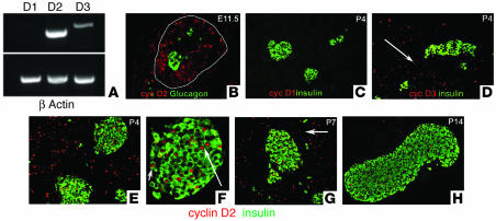
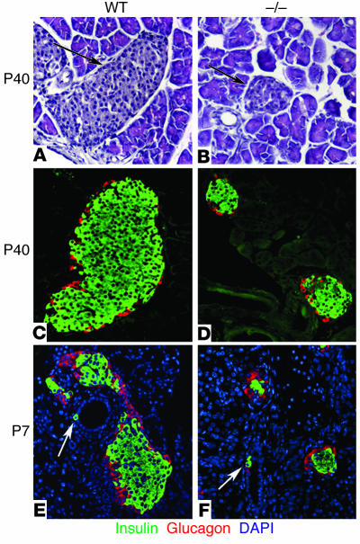
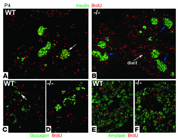
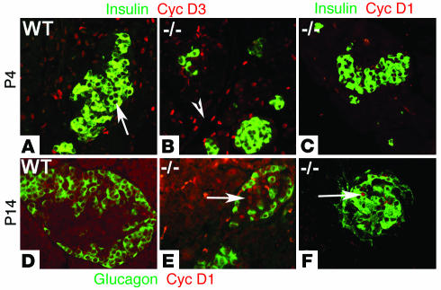
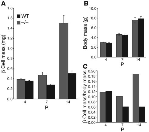
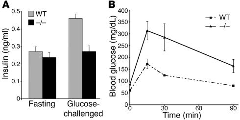
The endocrine pancreas undergoes major remodeling during neonatal development when replication of differentiated beta cells is the major mechanism by which beta cell mass is regulated. The molecular mechanisms that govern the replication of terminally differentiated beta cells are unclear. We show that during neonatal development, cyclin D2 expression in the endocrine pancreas coincides with the replication of endocrine cells and a massive increase in islet mass. Using cyclin D2-/- mice, we demonstrate that cyclin D2 is required for the replication of endocrine cells but is expendable for exocrine and ductal cell replication. As a result, 14-day-old cyclin D2-/- mice display dramatically smaller islets and a 4-fold reduction in beta cell mass in comparison to their WT littermates. Consistent with these morphological findings, the cyclin D2-/- mice are glucose intolerant. These results suggest that cyclin D2 plays a key role in regulating the transition of beta cells from quiescence to replication and may provide a target for the development of therapeutic strategies to induce expansion and/or regeneration of beta cells.


Figures

Figure 1
Figure 1
Cyclin D2 expression during pancreatic development. We carried out RT-PCR on RNA isolated from embryonic pancreata and stained pancreatic sections from various ages (as indicated) with anti_glucagon (green), anti_insulin (green), and anti_D-cyclin (red; as indicated) Ab’s. (A) Expression of cyclins D1, D2, and D3 in the developing pancreas by RT-PCR. RNA was isolated from E12.5 pancreata. (B) Cyclin D2 (cyc D2) expression in progenitor cells during early embryonic development. At E11.5, epithelial progenitor cells but not differentiated cells express cyclin D2. The dorsal pancreatic bud is outlined. At this stage, cyclin D2 expression is restricted to the epithelium and is not observed in the mesenchyme. (C) At P4, cyclin D1 (cyc D1) expression is not observed in the pancreas. (D) Cyclin D3 (cyc D3) is expressed in the exocrine and ductal (arrow) tissue but excluded from the endocrine pancreas. (E) Cyclin D2 is expressed throughout the pancreas at P4. Costaining with insulin Ab shows that cyclin D2 is expressed in the islets. At this stage, 34.5% ± 2.0% of islet cells express cyclin D2. (F) Higher magnification of an islet from E showing cyclin D2 expression is localized to the nucleus of β cells (arrows). (G) Cyclin D2 continues to be expressed in the endocrine pancreas at P7 (33.2% ± 4% of islet cells) and in the ductal tissue (arrow). (H) By P14, few cells in the endocrine pancreas express cyclin D2 (11.3% ± 1.4% of islet cells). Exocrine and ductal tissues do not show any expression of cyclin D2 at this stage.
Figure 2
Figure 2
Islet morphology and neogenesis of endocrine cells in WT and cyclin D2–/– mice. We stained pancreatic sections from 6-week-old WT and cyclin D2–/– (_/_) littermates with anti_glucagon and anti_insulin Ab’s. We estimated the islet size by measuring the longest axis of ten randomly selected islets that were at least ten cells wide. (A and B) H&E-stained pancreata from WT (A) and cyclin D2–/– (B) mice. The densely nucleated islets are indicated by arrows. (C) WT mice display a characteristic structure with α cells on the periphery and β cells at the core of the islet. (D) The cyclin D2–/– mice displayed dramatically smaller islets, although the morphology of α cells on the periphery and β cells at the core of the islet are preserved. The mean islet diameter of WT mice is 202 ± 13 μm versus 76.5 ± 4 μm for the cyclin D2–/– mice (P < 0.0001). (E and F) Pancreatic sections from P7 WT and cyclin D2–/– littermates stained with anti_insulin and anti_glucagon Ab’s. DAPI nuclear stain allows for the easy identification of ductal cells based on their characteristic morphology. Insulin-positive ductal cells in WT (E) and in cyclin D2–/– (F) littermates are shown (arrows). The frequency of hormone-positive ductal cells in the cyclin D2–/– mice is similar to that of WT littermates.
Figure 3
Figure 3
Incorporation of BrdU during postnatal pancreatic development of WT and cyclin D2–/– mice. BrdU was injected in 4- and 7-day-old mice 2 hours before being sacrificed. (A and B) Sections from pancreata costained with anti_insulin and anti_BrdU Ab’s. (A) In P4 WT mice, a fraction of β cells that incorporate BrdU are evident during the first week of postnatal development. Quantification of 20 representative islets showed that 9.2% of β cells incorporated BrdU in P4 WT mice. Arrow indicates an example of an islet with BrdU-positive β cells. (B) In cyclin D2–/– littermates, β cells that incorporated BrdU are not observed. Arrows indicate islets in cyclin D2–/– that do not contain BrdU-positive β cells. (C and D) Sections from pancreas costained with anti_glucagon and anti_BrdU Ab’s. (C) Glucagon-positive cells incorporate BrdU in the WT pancreas. Arrow indicates an example of an islet with a BrdU-positive α cell. (D) No glucagon-positive cells that incorporate BrdU are observed. (E and F) Sections from pancreas costained with anti_amylase and anti_BrdU Ab’s. BrdU incorporation is similar in exocrine and ductal tissue of the pancreata from WT (E) and cyclin D2–/– mice (F).
Figure 4
Figure 4
Expression of cyclin D1 and D3 in WT and cyclin D2–/– mice during postnatal development. (A and B) Sections from pancreata of 4-day-old WT and cyclin D2–/– mice were stained with anti_cyclin D3 and anti_insulin Ab’s. (A) In P4 WT mice, cyclin D3 expression is observed in exocrine and ductal tissue but is not observed in the endocrine pancreas. Arrow indicates islet lacking cyclin D3 expression. (B) In cyclin D2–/– littermates, cyclin D3 expression is observed in exocrine and ductal tissue (arrowhead) but not observed in the β cells. (C) Sections from pancreata of 4-day-old cyclin D2–/– mice were stained with anti_cyclin D1 and anti_insulin Ab’s. Cyclin D1 expression is not observed in the β cells in cyclin D2–/– mice. (D_F) Sections from pancreata of 14-day-old WT and cyclin D2–/– mice are stained with anti_cyclin D1, anti_glucagon (D and E), and anti_insulin (F) Ab’s. (D) Cyclin D1 is not expressed in the pancreas from WT P14 mice. (E) Cyclin D1 expression is observed within islets in pancreata from cyclin D2–/– littermates. Arrow indicates cyclin D1 expression within an islet of P14 cyclin D2–/– pancreas. (F) Cyclin D1 expression is observed in the nuclei of insulin-positive cells. Arrow indicates cyclin D1 expression in a β cell of P14 cyclin D2–/– pancreas.
Figure 5
Figure 5
β cell mass in WT and cyclin D2–/– mice during postnatal development. (A) The β cell mass per pancreas was estimated as the product of the relative cross-sectional area of β cells (determined by quantification of the cross-sectional area occupied by β cells divided by the cross-sectional area of total tissue) and the weight of the pancreas. β cell mass was measured at a postnatal age as indicated. Data are mean values ± SEM of four mice per genotype. In WT mice, β cell mass measurement showed a 4-fold increase by P14. In contrast, cyclin D2–/– littermates did not show any significant increase in β cell mass in the same period. (B) Body weights of cyclin D2 litters were measured to correspond to the β cell mass measurements during the postnatal period. The cyclin D2–/– mice weights do not differ from their littermates. (C) The relative β cell mass is calculated as a ratio of the β cell mass to body weight. Data are mean values ± SEM of ten mice per genotype.
Figure 6
Figure 6
Cyclin D2 mutant mice have decreased plasma insulin levels and impaired glucose tolerance. (A) Plasma insulin levels in WT and cyclin D2–/– mice following overnight fasting and 30 minutes after glucose challenge (2 g/kg body weight). Results are expressed as mean values ± SEM of four WT and cyclin D2–/– mice. (B) Blood glucose levels were measured after 16-hour fasting (time 0) and after glucose injection (2 g/kg body weight) in 12-week-old WT and cyclin D2–/– mice. Results are expressed as mean values ± SEM of three WT and cyclin D2–/– mice.

References

    1. Bruning JC, Winnay J, Cheatham B, Kahn CR. Differential signaling by insulin receptor substrate 1 (IRS-1) and IRS-2 in IRS-1-deficient cells. Mol. Cell. Biol. 1997;17:1513–1521. - PMC - PubMed
    1. Accili D. A kinase in the life of the β cell. J. Clin. Invest. 2001;108:1575–1576. doi:10.1172/JCI200114454. - PMC - PubMed
    1. Bonner-Weir S. Islet growth and development in the adult. J. Mol. Endocrinol. 2000;24:297–302. - PubMed
    1. Lingohr MK, Buettner R, Rhodes CJ. Pancreatic beta-cell growth and survival--a role in obesity-linked type 2 diabetes? [review] Trends Mol. Med. 2002;8:375–384. - PubMed
    1. Butler AE, et al. Beta-cell deficit and increased beta-cell apoptosis in humans with type 2 diabetes. Diabetes. 2003;52:102–110. - PubMed

Publication types