. 2004 Oct;114(7):969-78.
doi: 10.1172/JCI22103.

Prevention of type 1 diabetes by gene therapy

Affiliations

Prevention of type 1 diabetes by gene therapy

Chaorui Tian et al. J Clin Invest. 2004 Oct.

Abstract

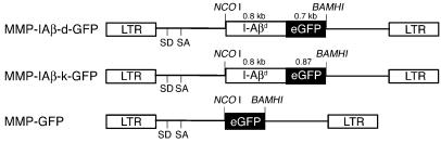
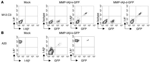
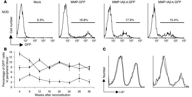
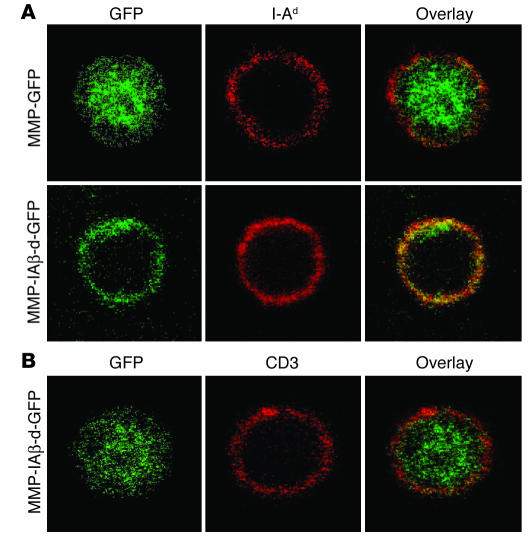
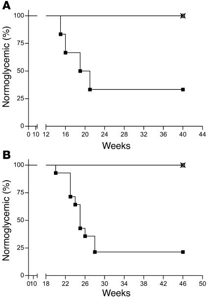
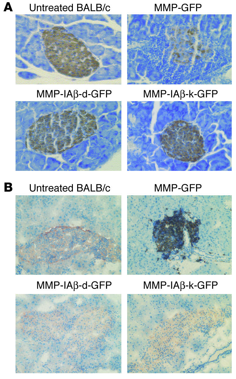
The autoimmune disease type 1 diabetes in humans and NOD mice is determined by multiple genetic factors, among the strongest of which is the inheritance of diabetes-permissive MHC class II alleles associated with susceptibility to disease. Here we examined whether expression of MHC class II alleles associated with resistance to disease could be used to prevent the occurrence of diabetes. Expression of diabetes-resistant MHC class II I-Abeta chain molecules in NOD mice following retroviral transduction of autologous bone marrow hematopoietic stem cells prevented the development of autoreactive T cells by intrathymic deletion and protected the mice from the development of insulitis and diabetes. These data suggest that type 1 diabetes could be prevented in individuals expressing MHC alleles associated with susceptibility to disease by restoration of protective MHC class II expression through genetic engineering of hematopoietic stem cells.


Figures

Figure 1
Figure 1
Diagram of MMP-based retroviral vectors encoding I-Aβd (MMP-IAβ-d-GFP) and I-Aβk (MMP-IAβ-k-GFP) fused to GFP; and diagram of the control MMP-GFP retroviral vector. LTR, long-terminal repeat.
Figure 2
Figure 2
Expression of retrovirally transduced MHC class II_GFP fusion proteins. (A) MMP-IAβ-d-GFP and MMP-IAβ-k-GFP retroviruses restore surface expression of MHC class II in I-Aβ chain_deficient M12.C3 cells. M12.C3 cells were cultured with media alone (mock) or infected with cell-free MMP-IAβ-k-GFP or MMP-IAβ-d-GFP retroviral supernatants for 3 days. Cells were harvested, and surface expression of MHC class II was analyzed by staining with anti-MHC mAb’s followed by flow cytometry. Shown is 1 representative example of 3 independent experiments. (B) I-Aβ chains encoded by MMP-IAβ-k-GFP are expressed on the surface of cells that express endogenous MHC class II. A20 cells (H-2d) were cultured with either media alone (mock) or cell-free MMP-IAβ-k-GFP viral supernatant for 3 days. Cells were harvested, and surface expression of MHC class II was analyzed by surface staining with anti_I-Aβk (10-3.6) and anti_I-Aβd (39–10–8) antibodies followed by flow cytometry. Shown is 1 representative example of 3 independent experiments.
Figure 3
Figure 3
Expression of retrovirally transduced MHC class II β chains in vivo. (A) MMP-IAβ-d-GFP and MMP-IAβ-k-GFP viruses effectively infect primary bone marrow cells. Bone marrow cells were harvested from 5-fluorouracil_treated NOD mice and transduced as described. Immediately after transduction, bone marrow cells were harvested and examined for GFP expression by flow cytometry. One representative experiment of 5 independent experiments is shown. (B) Expression of retrovirally transduced I-Aβ chains is stable. Expression of GFP in PBMCs of NOD mice reconstituted with MMP-IAβ-d-GFP_transduced (squares), MMP-IAβ-k-GFP_transduced (triangles), or control MMP-GFP_transduced bone marrow (diamonds), determined by flow cytometry. Shown are the mean values obtained for 5_6 mice per time point. The data are representative of 3 independent experiments. (C) MHC class II expression levels on the surface of PBMCs. Twenty weeks after bone marrow transplantation, PBMCs were harvested from mice reconstituted with bone marrow transduced with either MMP-IAβ-k-GFP (solid line) or control MMP-GFP virus (dashed line). Cells were stained with anti_I-Ad antibody 39-10-8, which recognizes all cell surface MHC class II, and analyzed by flow cytometry. Left panel: Total cells. Right panel: GFP+ transduced cells.
Figure 4
Figure 4
Subcellular localization of retrovirally encoded I-Aβ chains. (A) Retrovirally encoded I-Aβ chains are expressed on the surface of MHC class II_positive cells. PBMCs were harvested from NOD mice reconstituted with either MMP-GFP_transduced (top panels) or MMP-IAβ-d-GFP_transduced bone marrow (bottom panels), fixed, and stained with antibodies specific for GFP (left panels, green) and I-Ad (middle panels, red). The anti_I-Ad antibody used cross-reacts with I-Ag7. Right panels: Overlay images showing colocalization of retrovirally encoded I-Ad_GFP fusion proteins with endogenous I-Ag7 on the surface in yellow. (B) Retrovirally encoded I-Aβ chains are expressed in the cytoplasm of surface-MHC class II negative cells. PBMCs from NOD mice reconstituted with MMP-IAβ-d-GFP_transduced bone marrow were fixed and stained with antibodies specific for GFP (left panel, green) and anti-CD3 (middle panel, red). An overlay of the 2 images is shown in the right panel, demonstrating that I-Aβd_GFP does not colocalize with CD3 on the cell surface of MHC class II_negative cells. Shown are representative results of 3 independent experiments.
Figure 5
Figure 5
NOD mice reconstituted with either MMP-IAβ-d-GFP_ or MMP-IAβ-k-GFP_transduced bone marrow are protected from diabetes. (A) NOD mice were reconstituted with bone marrow retrovirally transduced with MMP-IAβ-d-GFP (open circles, n = 6), MMP-IAβ-k-GFP (×, n = 6), or control MMP-GFP (filled squares, n = 6). Shown are the percentages of normoglycemic mice at each time point after bone marrow transplantation. (B) NOD mice reconstituted with bone marrow retrovirally transduced with either MMP-IAβ-d-GFP or MMP-IAβ-k-GFP are protected from cyclophosphamide-induced diabetes. NOD mice were reconstituted with bone marrow transduced with MMP-IAβ-d-GFP (open circles, n = 12), MMP-IAβ-k-GFP (×, n = 12), or control MMP-GFP (filled squares, n = 14). Twenty-one weeks after bone marrow transplantation, mice were injected intraperitoneally with 200 mg/kg cyclophosphamide. Shown are the percentages of normoglycemic mice at each time point after bone marrow transplantation. Data in A and B are the combined results of 2 independent experiments. In all experiments, blood glucose levels were measured weekly.
Figure 6
Figure 6
Prevention of insulitis in NOD mice reconstituted with MMP-IAβ-d-GFP_ or MMP-IAβ-k-GFP_transduced bone marrow. (A) NOD mice reconstituted with MMP-IAβ-d-GFP_, MMP-IAβ-k-GFP_, or MMP-GFP_transduced bone marrow were sacrificed 20 weeks after transplantation. Pancreata were then harvested, fixed, paraffin-embedded, sectioned, and stained with anti-insulin antibodies, and counterstained with hematoxylin. Islets from mice reconstituted with MMP-GFP_transduced bone marrow showed significant mononuclear cell infiltration, and decreased storage of insulin (brown staining, top right panel). In contrast, islets from mice reconstituted with either MMP-IAβ-d-GFP_transduced (bottom left) or MMP-IAβ-k-GFP_transduced bone marrow (bottom right) were free of cellular infiltration, and levels of insulin storage were comparable to those observed in tissue sections from control BALB/c pancreas (top left). Shown are representative sections from 3 independent experiments. (B) Cells infiltrating NOD islets are predominantly CD3+. NOD mice reconstituted with MMP-IAβ-d-GFP_, MMP-IAβ-k-GFP_, or MMP-GFP_transduced bone marrow were sacrificed 20 weeks after bone marrow transplantation. Pancreata were snap-frozen, sectioned, fixed, and stained with antibodies specific for insulin (red) and CD3 (black), and then counterstained with hematoxylin (blue). Pancreata from NOD mice reconstituted with MMP-GFP_transduced bone marrow (top right panel) show peri-islet infiltration of CD3+ cells, and little insulin staining. In contrast, pancreata from mice reconstituted with either MMP-IAβ-d-GFP_transduced (bottom left) or MMP-IAβ-k-GFP_transduced bone marrow (bottom right) had no visible cellular infiltrate, and insulin staining was comparable to that observed in control BALB/c pancreata (top left). Shown are representative sections from 3 independent experiments.
Figure 7
Figure 7
Expression of retrovirally transduced diabetes-resistant MHC class II genes in bone marrow_derived cells prevents the development of functional autoreactive T cells in NOD mice. Twenty to twenty-eight weeks after reconstitution, splenocytes were harvested from NOD mice reconstituted with MMP-IAβ-d-GFP_, MMP-IAβ-k-GFP_, or control MMP-GFP_transduced bone marrow. Single-cell suspensions were prepared and cultured overnight with whole insulin protein, GAD 65 peptide 206_220, or, as a control, peptide 85_99 of MBP. The frequency of cells producing IFN-γ (A), IL-2 (B), and IL-4 (C) was then measured by cytokine ELISPOT assays. Responses to GAD 65 or insulin by splenocytes from NOD mice reconstituted with MMP-GFP_transduced bone marrow (black bars) were compared with responses to GAD 65 or insulin by cells from NOD mice reconstituted with either MMP-IAβ-d-GFP_ or MMP-IAβ-k-GFP_transduced bone marrow (white bars). Shown is 1 representative experiment of 3.
Figure 8
Figure 8
Central deletion of autoreactive T cells is mediated by retrovirally encoded MHC class II β chain. Eleven weeks after bone marrow transplantation, NOD mice reconstituted with either MMP-IAβ-d-GFP_ or control MMP-GFP_transduced bone marrow were sacrificed, and single-cell thymocyte suspensions were prepared. Single-cell suspensions were also prepared from naive age-matched NOD and BALB/c control mice. Thymocytes were stained with anti-CD4 and anti-CD8 antibodies, annexin V, and I-Ag7 tetramers loaded with either the control CLIP peptide or BDC-15 peptide, and analyzed by flow cytometry. (A) Single-positive CD4 T cells from mice reconstituted with bone marrow transduced with MMP-IAβ-d-GFP do not bind I-Ag7/BDC-15 tetramers. Cells were gated on live annexin V_CD4+CD8_ single-positive CD4 T cells. The percentage of these cells that bound to I-Ag7/CLIP (top row) and I-Ag7/BDC-15 tetramers (bottom row) is shown in the upper right quadrant. Shown is 1 representative experiment of 3. (B) Specific deletion of I-Ag7/BDC-15_binding T cells in mice reconstituted with bone marrow transduced with MMP-IAβ-d-GFP. Shown is expression of CD4 and CD8 after gating on I-Ag7/BDC-15 tetramer+, annexin V_ (live) cells. The total number of CD4+ single-positive T cells that bound the I-Ag7/BDC-15 tetramer was calculated by multiplication of the frequency of these cells by the total number of thymocytes recovered and is shown in the upper left quadrant. The total number of CD4+CD8+ double-positive thymocytes that bound the I-Ag7/BDC-15 tetramer was calculated similarly and is shown in the upper right quadrant. One representative experiment of 3 is shown.

Comment in

References

    1. Wicker LS, Todd JA, Peterson LB. Genetic control of autoimmune diabetes in the NOD mouse. Annu. Rev. Immunol. 1995;13:179–200. - PubMed
    1. Nepom GT, Kwok WW. Molecular basis for HLA-DQ associations with IDDM. Diabetes. 1998;47:1177–1184. - PubMed
    1. McDevitt H. The role of MHC class II molecules in susceptibility and resistance to autoimmunity. Curr. Opin. Immunol. 1998;10:677–681. - PubMed
    1. Morel PA, Dorman JS, Todd JA, McDevitt HO, Trucco M. Aspartic acid at position 57 of the HLA-DQ beta chain protects against Type I diabetes: a family study. Proc. Natl. Acad. Sci. U. S. A. 1988;85:8111–8115. - PMC - PubMed
    1. Ilonen J, et al. Estimation of genetic risk for type 1 diabetes. Am. J. Med. Genet. 2002;115:30–36. - PubMed

Publication types

MeSH terms

Substances