. 2004 Oct 6:3:25.
doi: 10.1186/1476-4598-3-25.

Genetic alterations and in vivo tumorigenicity of neurospheres derived from an adult glioblastoma

Affiliations

Genetic alterations and in vivo tumorigenicity of neurospheres derived from an adult glioblastoma

Patrizia Tunici et al. Mol Cancer. .

Abstract

Pediatric brain tumors may originate from cells endowed with neural stem/precursor cell properties, growing in vitro as neurospheres. We have found that these cells can also be present in adult brain tumors and form highly infiltrating gliomas in the brain of immunodeficient mice. Neurospheres were grown from three adult brain tumors and two pediatric gliomas. Differentiation of the neurospheres from one adult glioblastoma decreased nestin expression and increased that of glial and neuronal markers. Loss of heterozygosity of 10q and 9p was present in the original glioblastoma, in the neurospheres and in tumors grown into mice, suggesting that PTEN and CDKN2A alterations are key genetic events in tumor initiating cells with neural precursor properties.


Figures

Figure 1
Figure 1
Histological analysis of BT1 and BT1-derived tumors in nude mice. BT1 neurospheres (1 × 10e5) were stereotactically injected into the left hemisphere of nude mice (Charles River Italia, Calco, Italy; n = 3) or subcutaneously (n = 3). Nude mice were also injected with 1 × 10e5 BT1 adherent cells into the brain (n = 3) or subcutaneously (n = 3). Cells from BT2 were injected with similar procedures into nude mice. Control mice (n = 3) were injected with 1 × 10e5 neural stem/progenitor cells obtained from C57BL6J mice with previously described methods [11]. Fig 1A-B shows the GFAP staining in brown of coronal sections of the tumor derived from neurospheres (1A) or from adherent cells (1B). The right part on the figures correspond to the left hemisphere, were cells were injected. Fig. 1C-E show H-E staining of the primary tumor with features of a glioblastoma multiforme (1C) and of a tumor in mouse brain derived from neurospheres, showing an area with a prevailing aspect of oligodendroglioma (1D) or adherent cells, exhibiting anaplastic changes (1E). Fig. 1F-H show nestin staining of the primary tumor (1F) and of a tumor in mouse brain derived from neurospheres (1G) or adherent cells (1H).
Figure 2
Figure 2
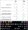
Genetic analysis on BT1, BT1-neurospheres and adherent cells and BT1-tumors in nude mice. DNA was extracted from frozen tissues, cell cultures or lymphocytes, using standard protocols. Primers, microsatellite markers and PCR conditions for LOH analysis were described before [12]. We also investigated markers 9S157 and 9S171 flanking the CDKN2A gene on 9p21. Before doing microsatellite analysis on mouse tumors we confirmed that PCR primers did not hybridize on mouse DNA. For cytogenetic analysis cells were harvested with 0.1 μg/ml Colcemid (Karyomax Colcemid, Life Technologies) overnight. Hypotonic treatment, fixation and GTG banding of metaphase chromosomes were performed with standard methods. The karyotypes were described in accordance with ISCN guidelines Spectral karyotyping was performed on metaphase cells according to the manufacturer's instructions (ASI, Carlsbad, CA) and to published procedures [13]. Spectral images were acquired and analyzed with an SD200 Spectral Bio-imaging System (ASI Ltd., MigdalHaemek, Israel) and a charged-coupled device camera (Hamamatsu, Bridgewater, NJ) connected to a Zeiss Axioskop 2 microscope (Carl Zeiss, Canada) and analyzed by the use of SKYVIEW (version 1.6.1; ASI) software. The upper panel shows the results of LOH analysis on 9p and 10q of the different samples outlined on the left. The lower panel illustrates a representative spectral karyotype of neurospheres obtained with the simultaneous hybridization of 24 combinatorially labeled chromosome painting probes. Karyotype display of chromosome banding (inverted DAPI) and SKY analysis (chromosomes were assigned a pseudo-color according to the measured spectrum) are shown. The number (7) next to the marker chromosome (der(3)) indicates the origin of inserted material.

References

    1. Singh SK, Clarke ID, Terasaki M, Bonn VE, Hawkins C, Squire J, Dirks PB. Identification of a cancer stem cell in human brain tumors. Cancer Res. 2003;63:5821–5828. - PubMed
    1. Hemmati HD, Nakano I, Lazareff JA, Masterman-Smith M, Geschwind DH, Bronner-Fraser M, Kornblum HI. Cancerous stem cells can arise from pediatric brain tumors. Proc Natl Acad Sci U S A. 2003;100:15178–15183. doi: 10.1073/pnas.2036535100. - DOI - PMC - PubMed
    1. Suslov ON, Kukekov VG, Ignatova TN, Steindler DA. Neural stem cell heterogeneity demonstrated by molecular phenotyping of clonal neurospheres. Proc Natl Acad Sci U S A. 2002;99:14506–14511. doi: 10.1073/pnas.212525299. - DOI - PMC - PubMed
    1. Li J, Yen C, Liaw D, Podsypanina K, Bose S, Wang SI, Puc J, Miliaresis C, Rodgers L, McCombie R, Bigner SH, Giovanella BC, Ittmann M, Tycko B, Hibshoosh H, Wigler MH, Parsons R. PTEN, a putative protein tyrosine phosphatase gene mutated in human brain, breast, and prostate cancer. Science. 1997;275:1943–1947. doi: 10.1126/science.275.5308.1943. - DOI - PubMed
    1. Chiariello E, Roz L, Albarosa R, Magnani I, Finocchiaro G. PTEN/MMAC1 mutations in primary glioblastomas and short-term cultures of malignant gliomas. Oncogene. 1998;16:541–545. doi: 10.1038/sj.onc.1201689. - DOI - PubMed

Publication types