. 2004 Dec;15(12):5616-22.
doi: 10.1091/mbc.e04-06-0454. Epub 2004 Oct 6.

Short tetracysteine tags to beta-tubulin demonstrate the significance of small labels for live cell imaging

Affiliations

Short tetracysteine tags to beta-tubulin demonstrate the significance of small labels for live cell imaging

Martin Andresen et al. Mol Biol Cell. 2004 Dec.

Abstract

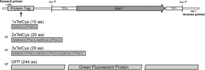
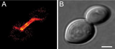
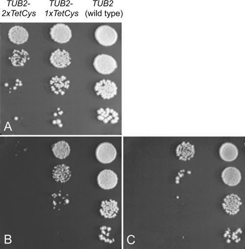
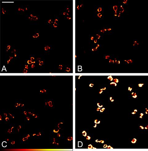
Genetically encoded tags are of fundamental importance for live cell imaging. We show that small tetracysteine (TetCys) tags can be highly advantageous for the functionality of the host protein compared with large fluorescent protein tags. One to three concatenated small TetCys tags as well as the large green fluorescent protein (GFP) were fused by integrative epitope tagging to the C terminus of beta-tubulin (Tub2) in the budding yeast Saccharomyces cerevisiae. The increasing tag size correlated with functional interference to the host protein. Tub2 tagged with either 1 x TetCys (10 amino acids [aa]) or 2 x TetCys (20 aa) was able to substitute Tub2 in haploid cells. In contrast, C-terminal tagging of Tub2 with 3 x TetCys (29 aa) or with GFP (244 aa) resulted in nonviable haploid cells. Cells expressing Tub2-1 x TetCys or Tub2-2 x TetCys were stained with FlAsH, which selectively binds to the TetCys-tag. The stained cells displayed dynamic FlAsH-labeled microtubules and low cellular background fluorescence. The presented approach to tag open reading frames (ORFs) at their native loci with very small TetCys-tags and the subsequent visualization of the tagged proteins in vivo can be extended in principle to any ORF in S. cerevisiae.


Figures

Figure 1.
Figure 1.
Schematic representation of the new modules in the plasmids pU1TetCys, pU2TetCys, pU3TetCys, and pUGFP. The four plasmids are identical except for the sequences of the epitopes. Therefore, the same set of oligonucleotides (forward primer and reverse primer) can be used to tag an ORF with any of these epitopes.
Figure 2.
Figure 2.
Tagging of TUB2 results in unique integration of modules into the genome. For the Southern blot, genomic DNA from diploid cells heterozygous for the tagged TUB2 gene was digested with AsnI and EcoRI, separated on a 1% agarose gel, and probed with a part of the kanMX-cassette. Lane 1, wild type; 2, strain expressing Tub2-1×TetCys; 3, strain expressing Tub2-2×TetCys; 4, strain expressing Tub2-3×TetCys; and 5, strain expressing Tub2-GFP. Expected fragment lengths are 1267 nucleotides (nt), 1297 nt, 1324 nt, and 1969 nt for lanes 2–5, respectively.
Figure 3.
Figure 3.
Tub2-GFP is integrated into microtubules when coexpressed with Tub2. Diploid heterozygous cells coexpressing Tub2 and Tub2-GFP were grown to logarithmic growth phase and imaged with a confocal microscope. (A) GFP-fluorescence. Displayed is a single optical plane. (B) Corresponding bright field image. Bar, 2 μm.
Figure 4.
Figure 4.
Growth phenotypes. Haploid wild-type and mutant strains expressing Tub2, Tub2-1×TetCys, or Tub2-2×TetCys, respectively, were grown overnight in glucose-containing medium. Then, 10-fold serial dilutions were spotted onto YPD-plates. The plates were incubated for 5 d at different temperatures. (A) 30°C. (B) 25°C. (C) 37°C.
Figure 5.
Figure 5.
FlAsH-labeled microtubules in live S. cerevisiae are dynamic, and the cells display low cellular background. Shown are haploid yeast cells expressing Tub2-1×TetCys or Tub2-2×TetCys without additional β-tubulin. Yeast cells were incubated overnight with FlAsH-EDT2 and subsequently imaged with a scanning confocal microscope. (A) Fluorescence image of cells expressing Tub2-1×TetCys. The majority of cells have stained microtubules. (B) Corresponding bright field image to A. Bar, 5 μm. (C) Fluorescence image of a budding haploid cell expressing Tub2-2×TetCys after staining with FlAsH-EDT2 overlaid with a bright field image. Bar, 2 μm. (D) Fluorescence intensity profile along the line indicated in C. The arrows indicate the position of the cell wall. (E) Time-lapse series of a cell expressing Tub2-1×TetCys after staining with FlAsH-EDT2. Confocal images were taken at the indicated time points. Displayed are maximum intensity projections of optical sections. The arrow points to a depolymerizing microtubule. (F) Corresponding bright field image to E. Bar, 5 μm.
Figure 6.
Figure 6.
Comparison of fluorescence intensities. Haploid cells expressing Atp1-1×TetCys (A), Atp1-2×TetCys (B), or Atp1-3×Tet-Cys (C) were grown to the logarithmic growth phase, stained with FlAsH-EDT2, and imaged with a confocal microscope. Haploid cells expressing Atp1-GFP were imaged under identical imaging conditions (D). The same look-up table is used for all images. Shown are single optical sections. Bar, 10 μm.

References

    1. Adams, S.R., Campbell, R.E., Gross, L.A., Martin, B.R., Walkup, G.K., Yao, Y., Llopis, J., and Tsien, R.Y. (2002). New biarsenical ligands and tetracysteine motifs for protein labeling in vitro and in vivo: synthesis and biological applications. J. Am. Chem. Soc. 124, 6063-6076. - PubMed
    1. Carminati, J.L., and Stearns, T. (1997). Microtubules orient the mitotic spindle in yeast through dynein-dependent interactions with the cell cortex. J. Cell Biol. 138, 629-641. - PMC - PubMed
    1. De Antoni, A., and Gallwitz, D. (2000). A novel multi-purpose cassette for repeated integrative epitope tagging of genes in Saccharomyces cerevisiae. Gene 246, 179-185. - PubMed
    1. Dolinski, K., et al. (2004). Saccharomyces Genome Database. http://www.yeastgenome.org/.
    1. Eggeling, C., Widengren, J., Rigler, R., and Seidel, C.A.M. (1999). Photostabilities of fluorescent dyes for single-molecule spectroscopy: mechanisms and experimental methods for estimating photobleaching in aqueous solution. In: Applied Fluorescence in Chemistry, Biology and Medicine, ed. W. Rettig, B. Strehmel, M. Schrader, and H. Seifert, Berlin: Springer, 193-240.

LinkOut - more resources