. 2004 Oct 6;24(40):8762-70.
doi: 10.1523/JNEUROSCI.2953-04.2004.

Characterization of the c-Jun N-terminal kinase-BimEL signaling pathway in neuronal apoptosis

Affiliations

Characterization of the c-Jun N-terminal kinase-BimEL signaling pathway in neuronal apoptosis

Esther B E Becker et al. J Neurosci. .

Erratum in

  • J Neurosci. 2004 Oct 27;24(43):1 p following 9722

Abstract

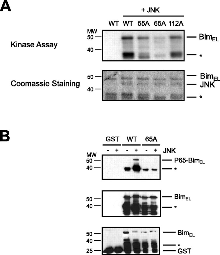
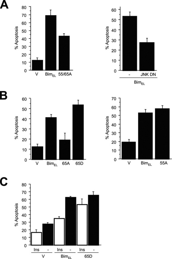
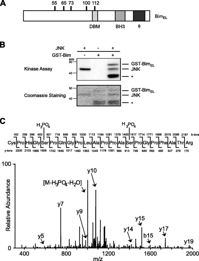
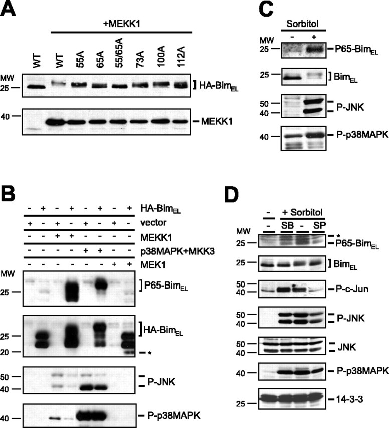
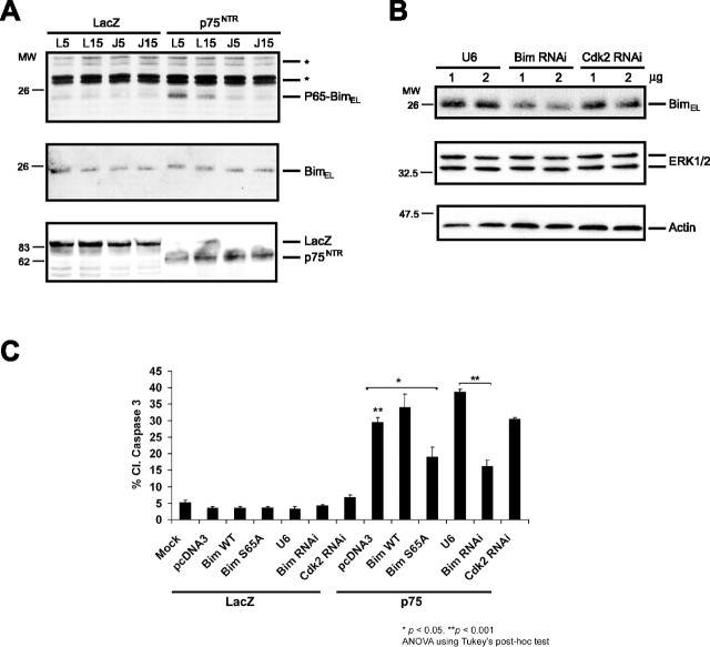
The c-Jun N-terminal kinase (JNK) signaling pathway plays a critical role in mediating apoptosis in the nervous system; however, the mechanisms by which JNK triggers neuronal apoptosis remain incompletely understood. Recent studies suggest that in addition to inducing transcription of pro-apoptotic genes, JNK also directly activates the cell death machinery. Here, we report that JNK catalyzed the phosphorylation of the BH3-only protein Bcl-2 interacting mediator of cell death (BimEL) at serine 65, both in vitro and in vivo. The JNK-induced phosphorylation of BimEL at serine 65 promoted the apoptotic effect of BimEL in primary cerebellar granule neurons. We also characterized the role of the JNK-BimEL signaling pathway in apoptosis that was triggered by overexpression of the p75 neurotrophin receptor (p75NTR). We found that activation of p75NTR induced the JNK-dependent phosphorylation of endogenous BimEL at serine 65 in cells. The genetic knockdown of BimEL by RNA interference or the expression of a dominant interfering form of BimEL significantly impaired the ability of activated p75NTR to induce apoptosis. Together, these results suggest that JNK-induced phosphorylation of BimEL at serine 65 mediates p75NTR-induced apoptosis. Our findings define a novel mechanism by which a death-receptor pathway directly activates the mitochondrial apoptotic machinery.


Figures

Figure 2.
Figure 2.
JNK phosphorylates BimEL at the distinct site of serine 65 in vitro. A, Recombinant GST-BimEL (WT) and recombinant BimEL phosphorylation mutants GST-BimELS55A (55A), GST-BimELS65A (65A), and GST-BimELT112A (112A) were incubated with no kinase or recombinant JNK1α1 and subjected to an in vitro kinase assay using [γ-32P]ATP. Phosphorylated substrates were separated by SDS-PAGE, stained with Coomassie blue (bottom panel), and analyzed by autoradiography (top panel). Degradation products of bacterially expressed GST-BimEL are indicated with an asterisk. B, Recombinant GST, GST-BimEL (WT), and GST-BimELS65A (65A) were subjected to an in vitro kinase assay with recombinant JNK1α1. Phosphorylated substrates were separated by SDS-PAGE and immunoblotted with the phospho65-Bim, Bim, or GST antibody. Degradation products of bacterially expressed proteins are marked with an asterisk.
Figure 4.
Figure 4.
Phosphorylation of BimEL at serine 65 promotes BimEL-mediated apoptosis. A, Cerebellar granule neurons were transfected with control vector (V), BimEL, or BimELS55/65A (55/65A) and β-galactosidase (left panel) or with control vector (V) or JNK DN together with BimEL and β-galactosidase (right panel). One day after transfection, cultures were switched from full medium to basal medium in the absence of survival factors. Cells were fixed after 8 hr and subjected to indirect immunofluorescence. Percentage of apoptosis is represented as mean ± SE. The expression of BimELS55/65A induced significantly less apoptosis than BimEL (n = 3; ANOVA; p < 0.0005). Expression of JNK DN significantly reduced BimEL-induced apoptosis (n = 3; ANOVA; p < 0.005). B, Cerebellar granule neurons were transfected with plasmids encoding nonspliceable cDNAs of BimEL, BimELS55A (55A), BimELS65A (65A), BimELS65D (65D), or control vector (V) and β-galactosidase. Cultures were treated as described above and analyzed by indirect immunofluorescence. The expression of BimELS65A induced significantly less apoptosis than BimEL and BimELS65D (mean ± SE; n = 4; ANOVA; p < 0.01 and p < 0.0005). The expression of BimELS55A did not reduce the apoptotic effect compared with wild-type BimEL (mean ± SE; n = 6; ANOVA; p = 0.4). C, Cerebellar granule neurons were transfected with plasmids encoding nonspliceable cDNAs of wild-type BimEL, BimELS65D (65D), or control vector (V) and β-galactosidase. One day after transfection, cultures were switched from full medium to basal medium in the presence (Ins) or absence (-) of insulin (10 μg/ml) to activate the IGF-I receptor. Cells were fixed after 8 hr and subjected to indirect immunofluorescence. Activation of the IGF-I receptor reduced Bim-mediated apoptosis (mean ± SE; n = 4; ANOVA; p < 0.0001) but had little effect on BimELS65D-induced apoptosis (p > 0.04).
Figure 1.
Figure 1.
JNK phosphorylates BimEL in vitro. A, The structure of BimEL is shown schematically. The location of the six putative proline-directed phosphorylation sites, the dynein binding motif (DBM), the BH3-only domain, and the hydrophobic domain (ϕ) are indicated. B, Recombinant GST-BimEL was incubated with recombinant JNK1α1 and subjected to an in vitro kinase assay using [γ-32P]ATP. Phosphorylated GST-BimEL was separated by SDS-PAGE, stained with Coomassie blue (bottom panel), and analyzed by autoradiography (top panel). Degradation products of bacterially expressed GST-BimEL are indicated with an asterisk. C, Representative tandem mass spectrum of one of the phosphopeptides (amino acids 51-71) derived from JNK-phosphorylated GST-BimEL. Fragment ions in the spectrum were sequenced from both the N and C termini (b- and y-type ions). Both serine 55 and serine 65 show additional mass from a phosphate residue.
Figure 3.
Figure 3.
JNK phosphorylates BimEL in vivo. A, HEK293T cells were transfected with 1 μg of wild-type BimEL (WT) or the BimEL phosphorylation mutants BimELS55A (55A), BimELS65A (65A), BimELS73A (73A), BimELS100A (100A), and BimELT112A (112A) alone or together with 0.5 μg of activated MEKK1. The electrophoretic mobility of the BimEL proteins was assessed by SDS-PAGE and immunoblotting with the HA antibody. B, HEK293T cells were transfected with 1 μg of wild-type BimEL or vector control alone or together with 0.5 μg of activated MEKK1, p38MAPK plus MKK3, or activated MEK1. Lysates of transfected cells were immunoblotted with the phospho65-Bim, Bim, phospho-JNK (P-JNK), or phospho-p38MAPK (P-p38MAPK) antibody. Untagged BimEL is indicated with an asterisk. C, HEK293T cells were treated with sorbitol (500 mm) for 3 hr. Lysates of cells were immunoblotted with the phospho65-Bim, Bim, phospho-JNK (P-JNK), or phospho-p38MAPK (P-p38MAPK) antibody. D, HEK293T cells were treated with sorbitol (500 mm) for 3 hr in the presence of vehicle or the p38MAPK and JNK inhibitors SB203580 (5 μm) and SP600125 (10 μm), respectively. Lysates of cells were immunoblotted with the phospho65-Bim, Bim, phospho-c-Jun (P-c-Jun), phospho-JNK (P-JNK), JNK, phospho-p38MAPK (P-p38MAPK), or 14-3-3 antibody. Aspecific bands recognized by the phospho65-Bim antibody are indicated with an asterisk.
Figure 5.
Figure 5.
p75NTR-induced apoptosis activates the JNK-dependent phosphorylation of BimEL and is dependent on BimEL. A, PC12 cells were infected with p75NTR or control adenovirus (LacZ) together with either 5 or 15 MOI of JBD-JIP (J) or control (L) adenovirus. Lysates were immunoblotted with phospho65-Bim or Bim antibody. Aspecific bands recognized by the phospho65-Bim antibody are indicated with an asterisk. B, Lysates of PC12 cells transfected with increasing amounts of control vector (U6), BimEL RNA interference plasmid (Bim RNAi), or control RNAi plasmid (Cdk2 RNAi) were immunoblotted with the Bim (top panel), p42 MAP kinase (ERK1/2; middle panel), or actin antibody (bottom panel). C, PC12rtta cells were transfected with the indicated plasmids and subsequently infected with p75NTR (p75) or control adenovirus (LacZ). Twenty-four hours after infection, cells were fixed and immunostained for cleaved caspase-3. Double asterisk indicates a difference of p < 0.001 between p75NTR-infected, pCDNA3-transfected cells (bar 8) and mock or LacZ-infected cells (bars 1-7), and p75NTR-infected, U6-transfected cells (bar 11) and Bim RNAi-transfected cells (bar 12). Single asterisk indicates a difference of p < 0.05 between p75NTR-infected, pCDNA3-transfected cells (bar 8) and BimELS65A-transfected cells (bar 10), as determined by ANOVA.

References

    1. Bamji SX, Majdan M, Pozniak CD, Belliveau DJ, Aloyz R, Kohn J, Causing CG, Miller FD (1998) The p75 neurotrophin receptor mediates neuronal apoptosis and is essential for naturally occurring sympathetic neuron death. J Cell Biol 140: 911-923. - PMC - PubMed
    1. Bhakar AL, Howell JL, Paul CE, Salehi AH, Becker EB, Said F, Bonni A, Barker PA (2003) Apoptosis induced by p75NTR overexpression requires Jun kinase-dependent phosphorylation of BAD. J Neurosci 23: 11373-11381. - PMC - PubMed
    1. Coffey ET, Smiciene G, Hongisto V, Cao J, Brecht S, Herdegen T, Courtney MJ (2002) c-Jun N-terminal protein kinase (JNK) 2/3 is specifically activated by stress, mediating c-Jun activation, in the presence of constitutive JNK1 activity in cerebellar neurons. J Neurosci 22: 4335-4345. - PMC - PubMed
    1. Dechant G, Barde YA (2002) The neurotrophin receptor p75(NTR): novel functions and implications for diseases of the nervous system. Nat Neurosci 5: 1131-1136. - PubMed
    1. D'Mello SR, Galli C, Ciotti T, Calissano P (1993) Induction of apoptosis in cerebellar granule neurons by low potassium: inhibition of death by insulin-like growth factor I and cAMP. Proc Natl Acad Sci USA 90: 10989-10993. - PMC - PubMed

Publication types

MeSH terms

Substances