. 2004 Oct 6;24(40):8806-17.
doi: 10.1523/JNEUROSCI.2726-04.2004.

Distribution and injury-induced plasticity of cadherins in relationship to identified synaptic circuitry in adult rat spinal cord

Affiliations

Distribution and injury-induced plasticity of cadherins in relationship to identified synaptic circuitry in adult rat spinal cord

John H Brock et al. J Neurosci. .

Abstract

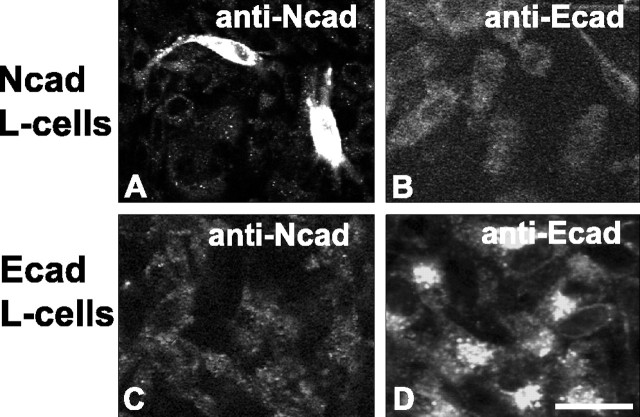
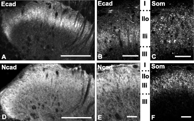
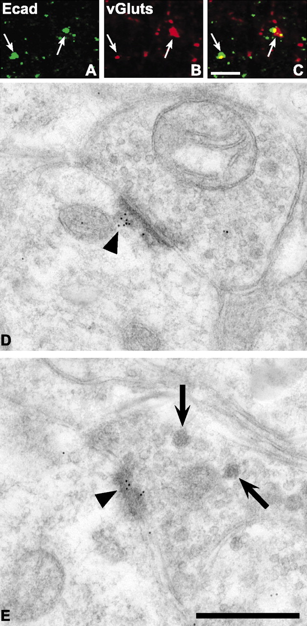
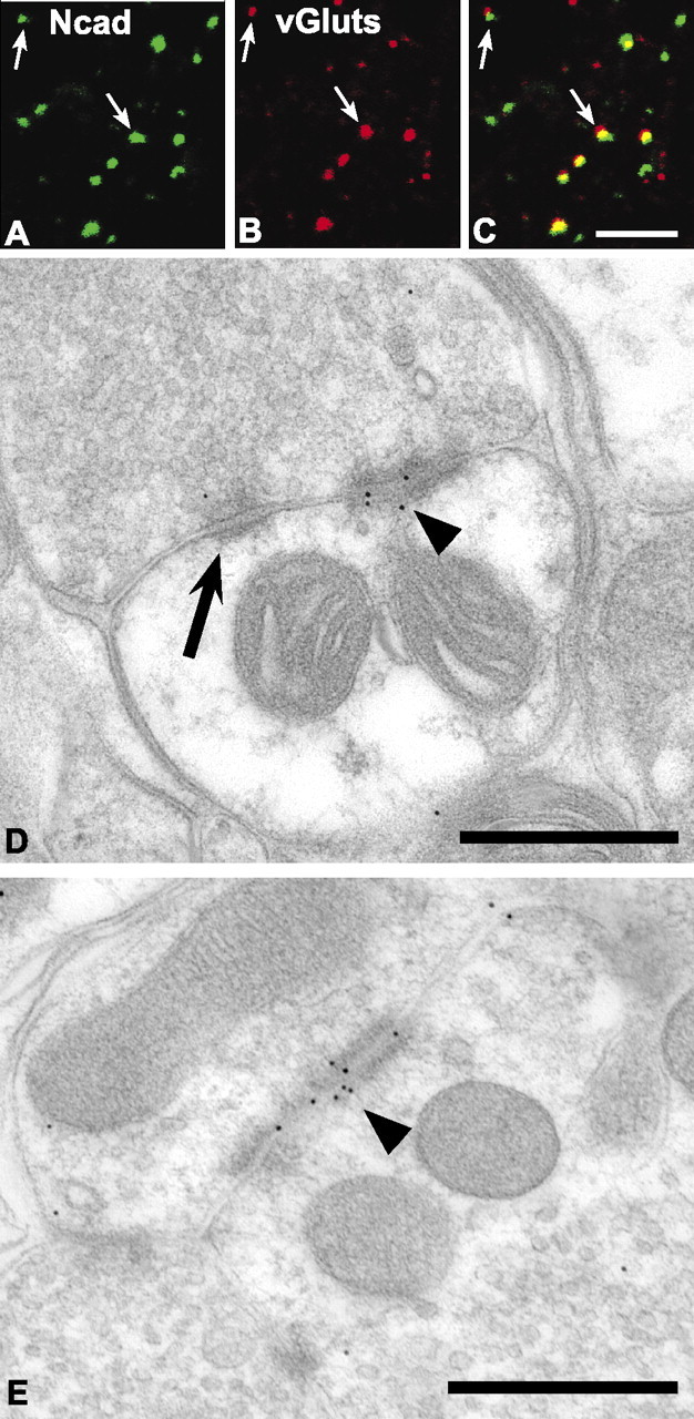
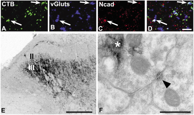
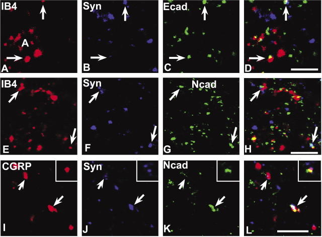
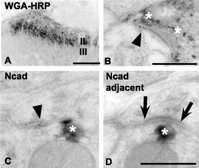
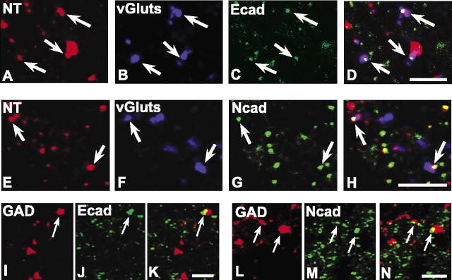
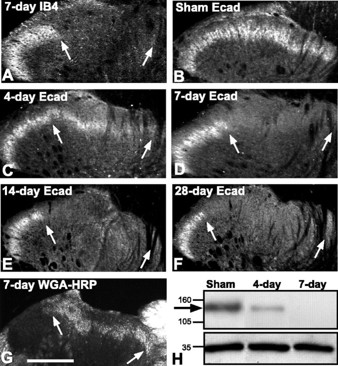
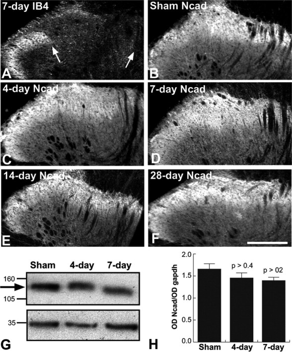
Cadherins are synaptically enriched cell adhesion and signaling molecules. In brain, they function in axon targeting and synaptic plasticity. In adult spinal cord, their localization, synaptic affiliation, and role in injury-related plasticity are mostly unexplored. Here, we demonstrate in adult rat dorsal horn that E- and N-cadherin display unique patterns of localization to functionally distinct types of synapses of intrinsic and primary afferent origin. Within the nociceptive afferent pathway to lamina II, nonpeptidergic C-fiber synapses in the deeper half of lamina II (IIi) contain E-cadherin but mostly lack N-cadherin, whereas the majority of the peptidergic C-fiber synapses in the outer half of lamina II (IIo) contain N-cadherin but lack E-cadherin. Approximately one-half of the Abeta-fiber terminations in lamina III contain N-cadherin; none contain E-cadherin. Strikingly, the distribution and levels of these cadherins are differentially affected by sciatic nerve axotomy, a model of neuropathic pain in which degenerative and regenerative structural plasticity has been implicated. Within the first 7 d after axotomy, E-cadherin is rapidly and completely lost from the dorsal horn synapses with which it is affiliated, whereas N-cadherin localization and levels are unchanged; such patterns persist through 28 d postlesion. The loss of E-cadherin thus occurs before the onset of mechanical hyperalgesia (approximately 10-21 d postlesion), as reported previously. Together, the synaptic specificity displayed by these cadherins, coupled with their differential response to injury, suggests that they may proactively contribute to the maintenance of some, and incipient dismantling of other, synaptic circuits in response to nerve injury. Speculatively, such changes may ultimately contribute to subsequently emerging abnormalities in pain perception.


Figures

Figure 1.
Figure 1.
Specificity of N-cadherin and E-cadherin antibodies. L-cells expressing N-cadherin (A, B) or E-cadherin (C, D) were incubated with mouse anti-N-cadherin (A, C) or anti-E-cadherin (B, D) antibodies. The anti-N-cadherin antibody labels N-cadherin L-cells (A) but does not label E-cadherin-expressing L-cells (C); conversely, the anti-E-cadherin antibody labels E-cadherin L-cells (D) but does not label N-cadherin-expressing L-cells (B). Scale bar, 10 μm.
Figure 2.
Figure 2.
Differential laminar distribution patterns of E- and N-cadherin immunolabeling in dorsal horn. E-cadherin labeling is confined to a dense, superficial band (A) corresponding to lamina IIi (B), determined by comparison with the lamina II marker somatostatin (Som; C, F). N-cadherin immunolabeling is broader (D) and is more homogeneously distributed throughout all dorsal horn laminas (E). In this and subsequent figures, Roman numerals correspond to dorsal horn lamina. IIo, Lamina II outer; IIi, lamina II inner. In all images, medial is to the right. Scale bars: A, D, 100 μm; B, C, E, F, 50 μm.
Figure 3.
Figure 3.
E-cadherin localizes to excitatory synaptic junctions within lamina IIi of the adult dorsal horn. Confocal images showing puncta immunolabeled for E-cadherin (A; green) and vGluts (B; red), separately and as an overlay (C; regions of yellow indicate codistribution). Arrows indicate identical puncta in each set of separate and overlaid images. D, E, Electron micrographs showing immunogold localization of E-cadherin antibody to asymmetric postsynaptic densities (arrowheads). Large dense-core vesicles (arrows) can be seen in the terminal shown in E. Scale bars: A-C, 5 μm; D, E, 500 nm.
Figure 4.
Figure 4.
N-cadherin localizes to excitatory synaptic junctional complexes within lamina IIo of the adult dorsal horn. Confocal images showing puncta immunolabeled for N-cadherin (A; green) and vGluts (B; red), separately and as an overlay (C; regions of yellow indicate codistribution). Arrows indicate identical puncta in each set of separate and overlaid images. D, E, Electron micrographs showing immunogold localization of rabbit N-cadherin antibody to a punctum adherens junction (D; arrowhead) adjacent to a conventional asymmetric postsynaptic density that lacks gold particles (D; arrow). N-cadherin antibody binding was also localized to some axoaxonic synapses (E; arrowhead). Scale bars: A-C, 5 μm; D, E, 500 nm.
Figure 7.
Figure 7.
N-cadherin localizes to Aβ-fiber synapses in lamina III. The terminations of Aβ-fibers were identified by transganglionic transport of CTB conjugates injected into the sciatic nerve. A-D, Confocal images showing puncta (arrows) containing CTB-Alexa 488 (A; green) immunolabeled for the synaptic marker vGluts (B; blue) and N-cadherin (C; red), separately and as an overlay (D; regions of white indicate codistribution of all three labels). E, Low-power, light microscopic image through lumber dorsal horn showing typical lamina III termination pattern of large-diameter, myelinated Aβ-fiber primary afferents revealed by CTB-HRP transport and TMB histochemistry. F, Electron micrograph of Aβ-fiber synapse in lamina III. Rabbit N-cadherin antibody binding is localized to an asymmetric synaptic contact (arrowhead). TMB reaction product (asterisk) is contained within the presynaptic terminal. Scale bars: A-D, 5 μm; E, 100 μm; F, 500 nm.
Figure 5.
Figure 5.
E- and N-cadherin codistribute with markers of primary afferent C-fiber synapses in lamina II. The nonpeptidergic C-fiber afferents to lamina IIi are labeled by IB4 binding (A, E), whereas the peptidergic C-fiber afferents to lamina IIo are labeled by CGRP (I); those forming synaptic terminals are identified by synaptophysin colabeling (B, F, J), and many of these contain E-cadherin (C) or N-cadherin (G, K), shown separately and in the overlays (D, H, L; regions of white indicate codistribution of all three labels). Insets in I-L show a representative example of a peptidergic C-fiber synaptic punctum containing N-cadherin. Scale bars, 5 μm.
Figure 6.
Figure 6.
Immunogold electron microscopic localization of E- and N-cadherin at synaptic junctions formed by WGA-HRP-identified primary afferents in lamina II. A, Low-power, light microscopic image through lumber dorsal horn showing typical lamina II termination pattern of small-diameter, myelinated, and unmyelinated primary afferents. TMB histochemistry was used to reveal such terminals after selective uptake and transganglionic transport of WGA-HRP injected into the sciatic nerve. Medial is to the right. B, Electron micrograph of C-fiber synapse in lamina IIi. E-cadherin antibody binding (arrowhead) is localized to an asymmetric postsynaptic density. The presynaptic terminal is identified as a small-diameter (nociceptive) afferent because it contains TMB reaction product (asterisks). C, D, Serial electron micrographs through a C-fiber synapse in lamina IIi immunogold-labeled for mouse N-cadherin antibody binding. Gold particles are found at perisynaptic puncta adherens (C; arrowhead) but are absent from the conventional asymmetric synaptic junction that is seen in the immediately adjacent section (Ncad adjacent; D; arrows). Asterisks in C and D indicate TMB reaction product contained in the afferent terminal. Scale bars: A, 100 μm; B-D, 500 nm.
Figure 8.
Figure 8.
E- and N-cadherin localize to excitatory and inhibitory synapses of intrinsic origin in lamina II. Confocal images of intrinsic excitatory synaptic puncta identified by labeling for NT (A, E; red), vGluts (B, F; blue), and E-cadherin (C; green) or N-cadherin (G; green) are shown separately and as overlays (D, H; regions of white indicate codistribution of all three markers). Confocal images of intrinsic inhibitory synaptic puncta identified by codistribution of labeling for GAD (I, L; red) and E-cadherin (J; green) or N-cadherin (M; green) are shown separately and as overlays (K, N; regions of yellow indicate codistribution). Scale bars, 5 μm.
Figure 9.
Figure 9.
Rapid and complete loss of E-cadherin after sciatic nerve axotomy. In all images, the region between arrows corresponds to the sciatic nerve termination zone in lamina IIi. A, IB4 binding is completely lost from the sciatic nerve termination zone by 7 d after axotomy but remains as expected more laterally in the region corresponding to saphenous nerve terminations. B-F, Fluorescence photomicrographs showing E-cadherin labeling in lamina IIi. A normal pattern of immunolabel is seen in the sham-operated control side (B). Intensity of immunolabel diminishes at 4 d after axotomy (C) and is undetectable in the sciatic nerve termination zone (arrows) by 7 d after axotomy (D). The loss of E-cadherin persists at 14 d (E) and at 28 d after axotomy (F). Sections shown in A, B, and D are from the same animal. G, Dark-field photomicrograph showing mostly normal pattern of TMB-reacted WGA-HRP terminal transport to lamina IIi after sciatic nerve axotomy. WGA-HRP was injected 4 d after axotomy and killed 3 d later. H, Representative E-cadherin immunoblot of extracts of lamina I/II taken from indicated conditions and days after sciatic nerve axotomy. The arrow indicates the position of bands corresponding to E-cadherin. E-cadherin levels are diminished by 4 d and completely absent by 7 d after axotomy, matching the post-axotomy patterns of immunolabeling. The bottom bands correspond to GAPDH, used as a loading control. Medial is to the right in A-G. Scale bars: A-G, 100 μm.
Figure 10.
Figure 10.
N-cadherin protein levels and localization unaltered by sciatic nerve axotomy. A, IB4 binding at 7 d after axotomy is completely absent from the sciatic nerve termination zone (between arrows), as expected. B-F, Fluorescence photomicrographs showing N-cadherin labeling throughout laminas I-III. Normal patterns and intensity of immunolabel are seen in sham-operated (B) and lesioned (C-F) animals at each post-axotomy time point examined (4, 7, 14, and 28 d). Sections shown in A, B, and D are from the same animal. Medial is to the right in A-F. G, Representative N-cadherin immunoblot of extracts of lamina I/II taken from indicated conditions and days after sciatic nerve axotomy. The arrow indicates the position of bands corresponding to N-cadherin. The bottom bands correspond to GAPDH, used as a loading control. H, Densitometric time course analysis shows N-cadherin protein levels do not change significantly with sciatic nerve axotomy. Data are means + SEM of ratios of N-cadherin band intensity to GAPDH band intensity (OD Ncad/OD gapdh). Each p value corresponds to comparison between relevant lesion group and sham control group. Scale bar, A-F, 100 μm.

References

    1. Alvarez FJ, Morris HR, Priestley JV (1991) Sub-populations of smaller diameter trigeminal primary afferent neurons defined by expression of calcitonin gene-related peptide and the cell surface oligosaccharide recognized by monoclonal antibody LA4. J Neurocytol 20: 716-731. - PubMed
    1. Arndt K, Nakagawa S, Takeichi M, Redies C (1998) Cadherin-defined segments and parasagittal cell ribbons in the developing chicken cerebellum. Mol Cell Neurosci 10: 211-228. - PubMed
    1. Arvidsson J, Ygge J, Grant G (1986) Cell loss in lumbar dorsal root ganglia and transganglionic degeneration after sciatic nerve resection in the rat. Brain Res 373: 15-21. - PubMed
    1. Bao L, Wang HF, Cai HJ, Tong YG, Jin SX, Lu YJ, Grant G, Hokfelt T, Zhang X (2002) Peripheral axotomy induces only very limited sprouting of coarse myelinated afferents into inner lamina II of rat spinal cord. Eur J Neurosci 16: 175-185. - PubMed
    1. Benson DL, Tanaka H (1998) N-cadherin redistribution during synaptogenesis in hippocampal neurons. J Neurosci 18: 6892-6904. - PMC - PubMed

Publication types