

doi: 10.1128/EC.3.5.1359-1362.2004.
Rapid production of gene replacement constructs and generation of a green fluorescent protein-tagged centromeric marker in Aspergillus nidulans
Affiliations
- PMID: 15470263
- PMCID: PMC522605
- DOI: 10.1128/EC.3.5.1359-1362.2004
Item in Clipboard
Rapid production of gene replacement constructs and generation of a green fluorescent protein-tagged centromeric marker in Aspergillus nidulans
Eukaryot Cell.
2004 Oct.
Abstract
A method to rapidly generate gene replacement constructs by fusion PCR is described for Aspergillus nidulans. The utility of the approach is demonstrated by green fluorescent protein (GFP) tagging of A. nidulans ndc80 to visualize centromeres through the cell cycle. The methodology makes possible large-scale GFP tagging, promoter swapping, and deletion analysis of A. nidulans.
Figures
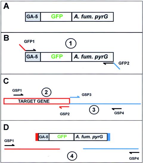
Construction of gene replacement constructs using fusion PCR. (A) Shown is the gene replacement cassette in which GA-5 is fused to GFP to act as a hinge between the target protein and GFP. The Af-pyrG gene (A. fum. pyrG) functions as a selectable nutritional marker. (B) The first PCR amplifies the GFP-pyrG cassette while incorporating 5′ and 3′ extensions complementary to 30 bp before and after the stop codon of the target gene with primers GFP1 and GFP2. (C) Two regions flanking the target gene stop codon are amplified with gene-specific primers 1 plus 2 and 3 plus 4 as indicated. (D) Fusion PCR is completed with the three amplified fragments and gene-specific primers 1 and 4.
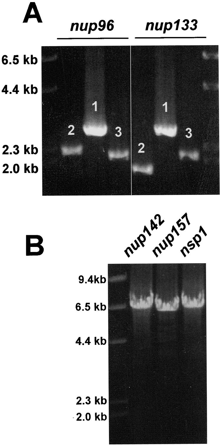
Generation of PCR fragments and a gene replacement construct. (A) Two examples of the three PCR fragments amplified for fusion PCR are shown. Fragments 1, 2, and 3 correspond to the fragments depicted in Fig. 1B and C. (B) Three examples of the final constructs generated by fusion PCR for GFP gene replacements.
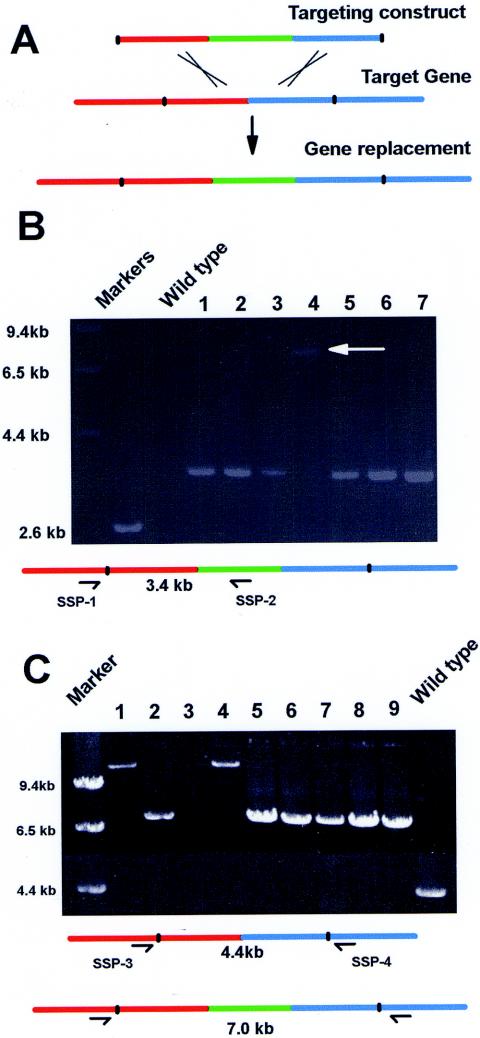
Analysis of a strain with a putative gene replacement by diagnostic PCR. (A) The targeting construct consists of 2 kb of sequence before the stop codon of the target gene (red) and the GFP-pyrG cassette (green) and 2 kb of sequence after the stop codon of the target gene (blue). After homologous recombination between the targeting domains, a gene replacement is generated with the target gene tagged at its C terminus by GFP. (B) One primer anchored outside the targeting sequence (SSP-1) and another within GFP (SSP-2) were used in PCRs containing wild-type DNA or DNA from seven strains transformed with a gene replacement construct. No amplified band is expected for the wild type, and a 3.4-kb band is expected for strains with replacement genes. The arrow indicates a band amplified from a transformant (lane 4) in which the linear replacement construct circularized before integration as explained in the text. (C) Two primers that anchored just outside the upstream (SSP-3) and downstream (SSP-4)targeting regions as shown were used to amplify DNA from nine transformants and a wild-type control. A 4.4-kb band is expected for the wild-type DNA PCR, which is replaced by a 7.0-kb band in strains with gene replacements. Larger amplified bands (lanes 1 and 4) or no bands (lane 3) indicate circularization of the targeting construct before integration, as explained in the text.
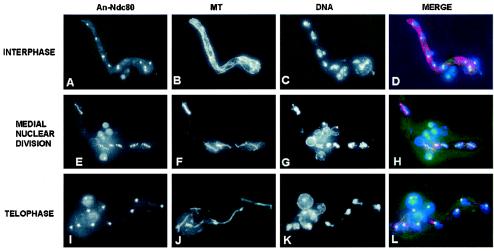
Location of the centromeric protein An-Ndc80 through the cell cycle. Interphase (A through D), medial mitotic (E through H), and telophase (I through L) cells are shown. The location of An-Ndc80-GFP (A, E, and I), microtubules (B, F, and J), DNA (C, G, and K), and a merge of all three (D, H, and L) during normal mitosis are shown. Notice the dramatic change in distribution of An-Ndc80 during the progression from interphase through mitosis.
References
-
- Bussink, H. J., and S. A. Osmani. 1999. A mitogen-activated protein kinase (MPKA) is involved in polarized growth in the filamentous fungus, Aspergillus nidulans. FEMS Microbiol. Lett. 173:117-125. - PubMed
-
- Fernandez-Abalos, J. M., H. Fox, C. Pitt, B. Wells, and J. H. Doonan. 1998. Plant-adapted green fluorescent protein is a versatile vital reporter for gene expression, protein localization and mitosis in the filamentous fungus, Aspergillus nidulans. Mol. Microbiol. 27:121-130. - PubMed
Publication types
MeSH terms
Substances
Grants and funding
LinkOut - more resources
Full Text Sources
Other Literature Sources
Molecular Biology Databases
