. 2004 Oct 27;23(21):4202-10.
doi: 10.1038/sj.emboj.7600391. Epub 2004 Oct 7.

Activation of IKKalpha target genes depends on recognition of specific kappaB binding sites by RelB:p52 dimers

Affiliations

Activation of IKKalpha target genes depends on recognition of specific kappaB binding sites by RelB:p52 dimers

Giuseppina Bonizzi et al. EMBO J. .

Abstract

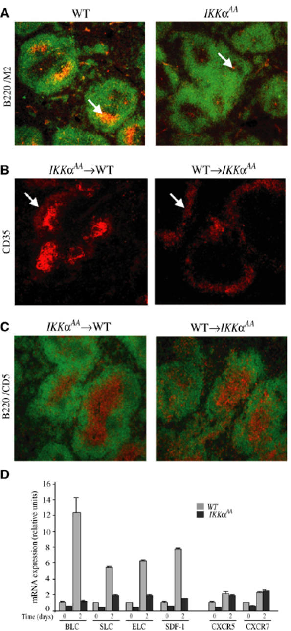
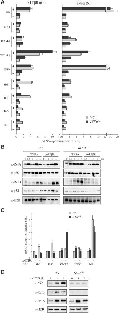
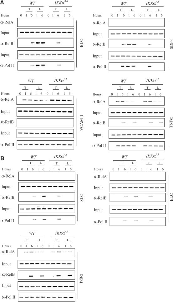
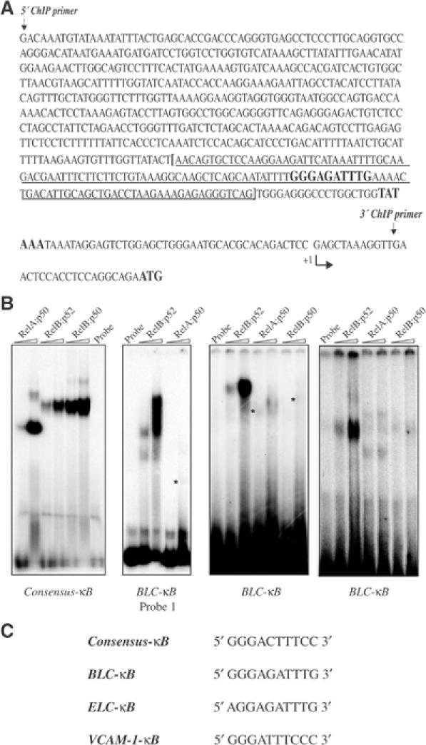
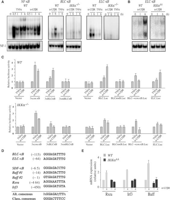
IkappaB Kinase (IKK)alpha is required for activation of an alternative NF-kappaB signaling pathway based on processing of the NF-kappaB2/p100 precursor protein, which associates with RelB in the cytoplasm. This pathway, which activates RelB:p52 dimers, is required for induction of several chemokine genes needed for organization of secondary lymphoid organs. We investigated the basis for the IKKalpha dependence of the induction of these genes in response to engagement of the lymphotoxin beta receptor (LTbetaR). Using chromatin immunoprecipitation, we found that the promoters of organogenic chemokine genes are recognized by RelB:p52 dimers and not by RelA:p50 dimers, the ubiquitous target for the classical NF-kappaB signaling pathway. We identified in the IKKalpha-dependent promoters a novel type of NF-kappaB-binding site that is preferentially recognized by RelB:p52 dimers. This site links induction of organogenic chemokines and other important regulatory molecules to activation of the alternative pathway.


Figures

Figure 1
Figure 1
Impaired FDC maturation and chemokine production in stromal cell-derived FDC requires IKKα. (A) Absence of mature FDC network in IkkαAA/AA mice. Cryosections of spleen from WT (n=6) and IkkαAA/AA (n=6) mice, isolated 7 days post-immunization with SRBC, were stained for FDCs (arrows) with FDC-M2 (orange) and anti-B220 (green). (B) Impaired FDC maturation is inherent to the IkkαAA/AA stroma. Lethally irradiated WT (n=6) or IkkαAA/AA (n=6) mice were reconstituted with IkkαAA/AA or WT bone marrow, respectively. Spleens were isolated 7 days after immunization with SRBC, cryosectioned and stained with anti-CD35. An FDC network is present in WT mice reconstituted with IkkαAA/AA bone marrow, while only perifollicular rings of CD35+ immature FDCs are present in IkkαAA/AA mice reconstituted with WT bone marrow. (C) Impaired B/T cell segregation in IkkαAA/AA spleens. Lethally irradiated WT (n=6) or IkkαAA/AA (n=6) mice reconstituted with IkkαAA/AA or WT bone marrow cells were immunized and analyzed as above, using anti-CD5 (to recognize T cells) and anti-B220 (to recognize B cells). Impaired B/T cell segregation is intrinsic to the IkkαAA/AA stroma. (D) Defective chemokine gene expression in IkkαAA/AA spleens. Total splenocytes from naïve and SRBC-immunized (day 2) WT (n=6) and IkkαAA/AA (n=6) mice were isolated. RNA was extracted and analyzed by RT–PCR for expression of mRNAs encoding BLC, SLC, ELC and SDF-1 and two of their receptors (CXCR5, CCR7). The results are averages±s.d. of three independent experiments normalized to the level of cyclophilin mRNA.
Figure 2
Figure 2
IKKα is required for LTβR-induced RelB:p52 nuclear translocation and chemokine expression in splenic stromal cells and myeloid dendritic cells. (A) IkkαAA/AA stromal cells and (C) BMDC exhibit specific defects in LTβR-induced gene expression. Total RNA was extracted from either WT or IkkαAA/AA stromal cells or BMDC before and after stimulation with 2 μg/ml agonistic anti-LTβR antibody or 20 ng/ml TNFα. Gene expression was analyzed by real-time PCR. Results are averages±s.d. of three independent experiments normalized to the level of cyclophilin mRNA. (B, D) Nuclear translocation of NF-κB proteins. Stromal cells (B) and BMDC (D) were stimulated with either anti-LTβR antibody or TNFα as indicated. At the indicated time points (h), nuclear extracts were prepared and analyzed by immunoblotting for the presence of the indicated NF-κB subunits. The levels of histone H2B were examined to control for loading and proper cell fractionation. Contamination with cytoplasmic proteins was monitored by blotting with anti-actin antibody (not shown).
Figure 3
Figure 3
IKKα is required for recruitment of RelB to the Blc, Sdf-1, Elc and Slc promoters. Primary cultures of stromal cells (A) and BMDC (B) from WT and IkkαAA/AA mice were left unstimulated or stimulated with TNFα (T) or anti-LTβR (L). At the indicated time points (h), the cells were collected and recruitment of RelA, RelB and the large subunit of RNA polymerase (Pol II) to the indicated promoter regions was examined by ChIP experiments.
Figure 4
Figure 4
The Blc and Elc promoters contain a unique κB site that is selectively recognized by RelB:p52 dimers. (A) The sequence of the 700 bp region, covering the proximal Blc promoter, contained within the ChIP primer set. The RelB-selective κB site and the TATA box are highlighted. The sequence contained within Probe 1 is indicated by the brackets and is underlined. (B) DNA-binding analysis. The different probes were incubated with two different amounts (250 and 500 ng) of the indicated NF-κB dimers and DNA binding was analyzed by EMSA. Note that the NF-κB subunits are not the full-length proteins, thus giving rise to complexes with different electrophoretic mobilities. (C) The sequences of the different κB sites used in these experiments.
Figure 5
Figure 5
Selective, IKKα-dependent activation of the Blc and Elc promoters by LTβR engagement and IKKα-dependent induction of Rxra, Irf3 and Baff mRNAs. (A, B) Engagement of LTβR selectively induces Blc-κB- and Elc-κB-binding activities. WT and IKKα-defective MEFs (A) and BMDC (B) were left unstimulated or stimulated with either TNFα or anti-LTβR for the indicated times. Nuclear extracts were prepared and incubated with 32P-labeled probes corresponding to the consensus κB site (NF-κB) or the Blc-κB and Elc-κB sites. DNA-binding activity was analyzed by EMSA. NF-1 DNA-binding activity was measured as an internal control. (C) Functional analysis of the different κB sites in the Blc and Elc promoters. Triple repeats of the consensus κB (conκB), Blc-κB and a mutant Blc-κB (mBlc-κB) sites were cloned upstream to a minimal SV40 promoter (pGL3-Promoter vector, Promega). In addition, the Blc (+12 to −688) and Elc (+530 to −320) promoter regions were cloned upstream to a luciferase reporter (pGL3-Basic vector, Promega). To determine the importance of the Blc-κB site, it was converted by site-directed mutagenesis either to an inactive mutant version (mκB) or the consensus κB (conκB) site. The different plasmids were transfected into WT and Ikkα−/− MEFs. After 6 h with TNFα or anti-LTβR, luciferase activity was determined. The results are averages±s.d. of three independent experiments normalized to β-galactosidase activity, produced by a cotransfected β-galactosidase expression vector. (D) Alignment of novel κB sites from the control regions of IKKα-dependent genes. The novel κB sites from the Blc, Elc and Sdf-1 5′ regulatory region were aligned with those identified by computer analysis in the regulatory regions of three other IKKα-dependent genes. These sites form a consensus sequence (Alt. consensus) that, although similar, is distinct from the one associated with the classical NF-κB pathway (Class. consensus). (E) Induction of Baff, Rxra and Irf3 is IKKα-dependent. Expression of the indicated mRNAs was analyzed by real-time PCR as described above, using RNA isolated from nonstimulated and anti-LTβR-stimulated stromal cells (Rxra and Irf3) and BMDCs (Baff) of the indicated genotypes.

References

    1. Alcamo E, Mizgerd JP, Horwitz BH, Bronson R, Beg AA, Scott M, Doerschuk CM, Hynes RO, Baltimore D (2001) Targeted mutation of TNF receptor I rescues the RelA-deficient mouse and reveals a critical role for NF-κB in leukocyte recruitment. J Immunol 167: 1592–1600 - PubMed
    1. Anest V, Hanson JL, Cogswell PC, Steinbrecher KA, Strahl BD, Baldwin AS (2003) A nucleosomal function for IκB kinase-α in NF-κB-dependent gene expression. Nature 423: 659–663 - PubMed
    1. Ansel KM, Cyster JG (2001) Chemokines in lymphopoiesis and lymphoid organ development. Curr Opin Immunol 13: 172–179 - PubMed
    1. Ansel KM, Ngo VN, Hyman PL, Luther SA, Forster R, Sedgwick JD, Browning JL, Lipp M, Cyster JG (2000) A chemokine-driven positive feedback loop organizes lymphoid follicles. Nature 406: 309–314 - PubMed
    1. Bonizzi G, Piette J, Schoonbroodt S, Greimers R, Havard L, Merville MP, Bours V (1999) Reactive oxygen intermediate-dependent NF-κB activation by interleukin-1β requires 5-lipoxygenase or NADPH oxidase activity. Mol Cell Biol 19: 1950–1960 - PMC - PubMed

Publication types

MeSH terms