. 2004 Dec 17;279(51):53643-52.
doi: 10.1074/jbc.M401438200. Epub 2004 Oct 7.

Characterization of the GRK2 binding site of Galphaq

Affiliations

Characterization of the GRK2 binding site of Galphaq

Peter W Day et al. J Biol Chem. .

Abstract

Heterotrimeric guanine nucleotide-binding proteins (G proteins) transmit signals from membrane bound G protein-coupled receptors (GPCRs) to intracellular effector proteins. The G(q) subfamily of Galpha subunits couples GPCR activation to the enzymatic activity of phospholipase C-beta (PLC-beta). Regulators of G protein signaling (RGS) proteins bind to activated Galpha subunits, including Galpha(q), and regulate Galpha signaling by acting as GTPase activating proteins (GAPs), increasing the rate of the intrinsic GTPase activity, or by acting as effector antagonists for Galpha subunits. GPCR kinases (GRKs) phosphorylate agonist-bound receptors in the first step of receptor desensitization. The amino termini of all GRKs contain an RGS homology (RH) domain, and binding of the GRK2 RH domain to Galpha(q) attenuates PLC-beta activity. The RH domain of GRK2 interacts with Galpha(q/11) through a novel Galpha binding surface termed the "C" site. Here, molecular modeling of the Galpha(q).GRK2 complex and site-directed mutagenesis of Galpha(q) were used to identify residues in Galpha(q) that interact with GRK2. The model identifies Pro(185) in Switch I of Galpha(q) as being at the crux of the interface, and mutation of this residue to lysine disrupts Galpha(q) binding to the GRK2-RH domain. Switch III also appears to play a role in GRK2 binding because the mutations Galpha(q)-V240A, Galpha(q)-D243A, both residues within Switch III, and Galpha(q)-Q152A, a residue that structurally supports Switch III, are defective in binding GRK2. Furthermore, GRK2-mediated inhibition of Galpha(q)-Q152A-R183C-stimulated inositol phosphate release is reduced in comparison to Galpha(q)-R183C. Interestingly, the model also predicts that residues in the helical domain of Galpha(q) interact with GRK2. In fact, the mutants Galpha(q)-K77A, Galpha(q)-L78D, Galpha(q)-Q81A, and Galpha(q)-R92A have reduced binding to the GRK2-RH domain. Finally, although the mutant Galpha(q)-T187K has greatly reduced binding to RGS2 and RGS4, it has little to no effect on binding to GRK2. Thus the RH domain A and C sites for Galpha(q) interaction rely on contacts with distinct regions and different Switch I residues in Galpha(q).


Figures

Figure 1
Figure 1
(A) Model of the Gαq-GRK2 RH domain complex and their interacting surfaces. The model of Gαq was homology modeled based on the AlF4 bound structure of Gαi in complex with RGS4 (11) and then docked with the RH domain as described in Experimental Procedures. The switch regions and the αA helix of Gαq (purple and yellow) are labeled, as are the α5 and α6 helices of the GRK2 RH domain. These structural elements constitute the principal interaction surfaces of each protein. The proposed plane of the plasma membrane runs along the top of the complex, as shown in the figure. The switch regions of Gαq are delineated by V182 to Y192 (switch I), V204- T224 (switch II), and D236-R247 (switch III). (B) Model of Gαq in complex with RGS4, based on the atomic structure of Gαi-RGS4 (11). The RH domains of GRK2 and RGS4 both interact with the switch regions of the G protein, but the surface of the RH domain used in the contact is unique. In the RGS4 complex, the α5 helix faces out of the page, while in the Gαq-GRK2 complex it forms the principal contact surface. Panels (C) and (D) represent views of Gαq and GRK2, respectively, as if the complex shown in panel (A) were opened like a book. (C) The GRK2-interacting surface of Gαq. The residues shown as ball-and-stick models with green carbons are those mutated and analyzed in this study. Thick circles indicate residues that had a dramatic effect upon mutation (as per Table 1), thin circles indicate an “intermediate” effect, and no circles indicate no effect, at least upon GRK2 binding and inhibition of IP3 release. The residues listed in orange are those that each Gαq residue is predicted to contact. The black sphere represents Mg2+. (D) The Gαq-interacting surface of the GRK2 RH domain. The residues shown as balland- stick models with green carbons are those mutated and analyzed in this (L118 and E130) and our previous study (22). Thick circles indicate residues that had a dramatic effect upon mutation, thin circles indicate an “intermediate” effect, and no circles indicate no effect, at least upon Gαq binding. The residues listed in purple are those that each GRK2 residue is predicted to contact. All panels were created using PyMOL (38). The coordinates of the model of the Gαq-GRK2 RH domain complex are available in a pdb file as Supplementary Data.
Figure 2
Figure 2
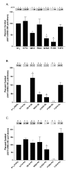
Interaction of GST-GRK2-(45–178) with Gαq point mutants activated by AlF4, the Q209L or the R183C mutation. (A) HEK-293 cells were transfected with EE tagged versions of Gαq point mutants and Gβ and Gγ constructs. Cells were lysed and binding to GST-GRK2-(45–178) in the presence (+) or the absence (−) of AlF4 was determined as described in Experimental Procedures. The (+) and (−) lanes represent 40% of the Gαq or Gαq mutant pulled down from the 125 μl of lysate. In these experiments we detect little to no binding of GST-GRK2-(45–178) to Gαq or Gαq point mutants in the absence of AlF4. Underneath the representative western blot the percent of each Gαq mutant pulled down by GST-GRK2-(45–178) in the presence of AlF4 is compared to the control, which is the percent of Gαq pulled down by GST-GRK2 (45–178) in the presence of AlF4, and is represented graphically as the percent of control + S.D. (B) HEK-293 cells were transfected with EE tagged versions of Gαq-Q209L point mutants and Gβ and Gγ constructs. Cells were lysed and binding of the QL mutants to GST-GRK2-(45–178) was determined as described in Experimental Procedures. Results are plotted as described in A. (C) HEK-293 cells were transfected with EE tagged versions of Gαq-R183C point mutants and Gβ and Gγ constructs. Cells were lysed and binding of the RC mutants to GST-GRK2-(45–178) was determined as described in Experimental Procedures. The lanes labeled “P” represent 40% of the Gαq or Gαq mutant that was present in the pulldown from 200 μl of lysate. The lanes in A, B and C labeled “L” represent 4% of total Gαq or Gαq mutant available in the lysate for pull-down. Results are plotted as described in A. The (⋆) indicates that the amount of the marked Gαq mutant pulled-down is significantly different (p < 0.05) by one-way ANOVA followed by a Dunnett post-test, than the amount of Gαq pulled-down by GST-GRK2. The (⋆⋆) indicates that statistical analysis could not be performed on the binding of GST-GRK2-RH to the K77A-QL, P185K-QL or P185K-RC mutants because there was no detectable pull-down. The data are averages from three to six independent experiments.
Figure 3
Figure 3
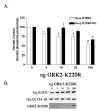
Effect of the Q152A point mutation in Gαq-RC on the ability of GRK2 to inhibit inositol phosphate production. (A) HEK-293 cells were transfected with 0.1 μg of the constitutively active Gαq-R183C or Gαq-Q152A-RC and 0.2 μg of myc, His-tagged Gβ and 0.1 μg of Gγ and increasing amounts of GRK2-K220R and empty vector up to a total of 1.0 μg of DNA. 24 hrs after transfection the cells were labeled with 2 μCi/ml myo-[3H]inositol and 16 hours later inositol phosphate production was determined, as described in Experimental Procedures. The results shown are averages from five independent experiments each done in triplicate and displayed as percent control ± S.D. The control is the inositol phosphate production stimulated by Gαq-R183C or Gαq- Q152A-RC in the absence of any co-expressed GRK2-K220R. A (⋆) denotes a statistically significant difference (p < 0.05) by two-way ANOVA followed by a Bonferroni post-test, between the indicated Gαq-Q152A-RC bar and the Gαq-RC bar transfected with the same amount of GRK2-K220R. A (#) indicates a statistically significant difference (p < 0.05) by one-way ANOVA followed by a Dunnett post-test, between the indicated bar and the control, either Gαq-RC or Gαq-Q152A-RC in the absence of cotransfected GRK2-K220R. (B) Western blots of total cellular lysates from a representative inositol phosphate experiment from (A) probed with the EE monoclonal antibody showing that increasing GRK2-K220R expression does not effect expression of Gαq-RC or Gαq-Q152A-RC. The bottom panel of Figure 4 B shows the level of GRK2- K220R overexpression. The bands corresponding to 10, 25 and 100 ng of GRK2-K220R transfected can be seen after very short exposures; however, the GRK2 band corresponding to 5 ng of cDNA transfected is barely visible, even after long exposures, suggesting that comparatively low levels of GRK2 expression can significantly inhibit Gαq signaling.
Figure 4
Figure 4
Interaction of GST-RGS2 with Gαq point mutants activated by AlF4, the Q209L or the R183C mutation. (A) HEK-293 cells were transfected with EE tagged versions of Gαq point mutants and Gβ and Gγ constructs. Cells were lysed and binding to GST-RGS2 in the presence (+) or the absence (−) of AlF4 was determined as described in Experimental Procedures. The (+) and (−) lanes represent 40% of the Gαq or Gαq mutant pulled down from the 125 μl of lysate. In these experiments we detect little to no binding of GST- RGS2 to Gαq or Gαq point mutants in the absence of AlF4. Underneath the representative western blot the percent of each Gαq mutant pulled down by GST- RGS2 in the presence of AlF4 is compared to the control, which is the percent of Gαq pulled down by GST-RGS2 in the presence of AlF4, and is represented graphically as the percent of control ± S.D. (B) HEK-293 cells were transfected with EE tagged versions of Gαq-Q209L point mutants and Gβ and Gγ constructs. Cells were lysed and binding of the QL mutants to GST-RGS2 was determined as described in Experimental Procedures. Results are plotted as described in A. (C) HEK-293 cells were transfected with EE tagged versions of Gαq-R183C point mutants and Gβ and Gγ constructs. Cells were lysed and binding of the RC mutants to GST-RGS2 was determined as described in Experimental Procedures. The lanes labeled “P” in B and C represent 40% of the Gαq or Gαq mutant that was present in the pull-down from 200 μl of lysate. The lanes in A, B and C labeled “L” represent 4% of total Gαq or Gαq mutant available in the lysate for pull-down. Results are plotted as described in A. The (⋆) indicates that the amount of the marked Gαq mutant pulled-down is significantly different (p < 0.05) by one-way ANOVA followed by a Dunnett post-test, than the amount of Gαq pulled-down by GST-RGS2. The (⋆⋆) indicates that statistical analysis could not be performed on the binding of GST-RGS2 to the K77A-QL, P185K-QL, P185K-RC, T187K or T187K-QL mutants because there was no detectable pull-down. The data are averages from three to six independent experiments.
Figure 5
Figure 5
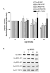
RGS2 inhibition of inositol phosphate production stimulated by Gαq-RC and Gαq-RC mutants. (A) HEK-293 cells were transfected with 0.1 μg of the constitutively active Gαq-R183C, Gαq-Q81A/RC, Gαq-R92A/RC, Gαq-Q152A/RC, or Gαq-RC/T187K and 0.2 μg of myc, His-tagged Gβ and 0.1 μg of Gγ and increasing amounts of RGS2 and empty vector up to a total of 1.0 μg of DNA. 24 hrs after transfection the cells were labeled with 2 μCi/ml myo-[3H]inositol and 16 hours later inositol phosphate production was determined, as described in Experimental Procedures. The results shown are averages from three independent experiments each done in triplicate and displayed as percent control + SD. The control is the inositol phosphate production stimulated by each mutant in the absence of any co-expressed RGS2. The statistical significance of the difference between the indicated bar and Gαq-R183C, in the absence of any additional mutations, transfected with equal amounts of RGS2 is denoted by ⋆ (p < 0.05) by oneway ANOVA followed by a Dunnett post-test. A (#) indicates a statistically significant difference (p < 0.05) by one-way ANOVA followed by a Dunnett post-test, between the indicated bar and the control, either Gαq-RC or a Gαq-RC mutant in the absence of cotransfected RGS2. (B) Western blots of total cellular lysates from a representative inositol phosphate experiment from (A) probed with the EE monoclonal antibody showing that increasing RGS2 expression does not effect expression of Gαq-RC or any of the mutants. We were not able to detect the level of RGS2 overexpression in these experiments; however the decrease in inositol phosphate production suggests that RGS2 expression was increased.
Figure 6
Figure 6
Further mapping of the Gαq/11 binding site on the GRK2 RH domain. Upper Panel. Glutathione-agarose beads bearing GST fusion proteins, either WT GSTGRK2-(45–178) or GST-GRK2-(45–178) substituted as indicated, were incubated with bovine brain extract (as a source of Gαq/11) in the presence (+) or absence (−) of aluminum fluoride (AlF4). Bound Gαq/11 was visualized by immunoblotting. Lower Panel. Fusion proteins used in the GST-pull-down assay above were separated by SDS-PAGE and visualized by Coomassie staining.

Similar articles

Cited by

References

    1. Bourne HR. Curr Opin Cell Biol. 1997;9:134–142. - PubMed
    1. Krupnick JG, Benovic JL. Annu Rev Pharmacol Toxicol. 1998;38:289–319. - PubMed
    1. Hausdorff WP, Caron MG, Lefkowitz RJ. FASEB J. 1990;4:2881–2889. - PubMed
    1. Goodman OB, Jr, Krupnick JG, Santini F, Gurevich VV, Penn RB, Gagnon AW, Keen JH, Benovic JL. Nature. 1996;383:447–450. - PubMed
    1. Ferguson SS, Downey WE, 3rd, Colapietro AM, Barak LS, Menard L, Caron MG. Science. 1996;271:363–366. - PubMed

Publication types

MeSH terms

LinkOut - more resources