. 2005 Jan;146(1):13-8.
doi: 10.1210/en.2004-1123. Epub 2004 Oct 7.

A system for studying epithelial-stromal interactions reveals distinct inductive abilities of stromal cells from benign prostatic hyperplasia and prostate cancer

Affiliations

A system for studying epithelial-stromal interactions reveals distinct inductive abilities of stromal cells from benign prostatic hyperplasia and prostate cancer

Wendy W Barclay et al. Endocrinology. 2005 Jan.

Abstract

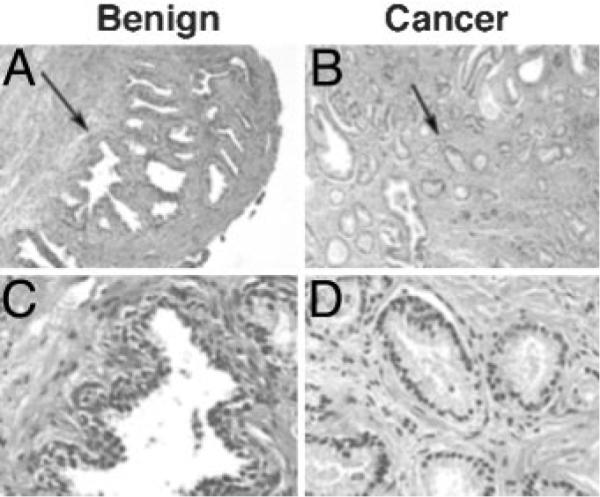
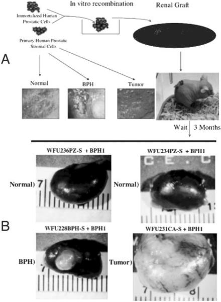
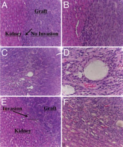
The development of normal and abnormal glandular structures in the prostate is controlled at the endocrine and paracrine levels by reciprocal interactions between epithelium and stroma. To study these processes, it is useful to have an efficient method of tissue acquisition for reproducible isolation of cells from defined histologies. Here we assessed the utility of a standardized system for acquisition and growth of prostatic cells from different regions of the prostate with different pathologies, and we compared the abilities of stromal cells from normal peripheral zone, benign prostatic hyperplasia (BPH-S), and cancer to induce the growth of a human prostatic epithelial cell line (BPH-1) in vivo. Using the tissue recombination method, we showed that grafting stromal cells (from any histology) alone or BPH-1 epithelial cells alone produced no visible grafts. Recombining stromal cells from normal peripheral zone with BPH-1 cells also produced no visible grafts (n = 15). Recombining BPH-S with BPH-1 cells generated small, well-organized, and sharply demarcated grafts approximately 3-4 mm in diameter (n = 9), demonstrating a moderate inductive ability of BPH-S. Recombining stromal cells from cancer with BPH-1 cells generated highly disorganized grafts that completely surrounded the host kidney and invaded into adjacent renal tissue, demonstrating induction of an aggressive phenotype. We conclude that acquisition of tissue from toluidine blue dye-stained specimens is an efficient method to generate high-quality epithelial and/or stromal cultures. Stromal cells derived by this method from areas of BPH and cancer induce epithelial cell growth in vivo, which mimics the natural history of these diseases.


Figures

Fig. 1
Fig. 1
Acquisition and culture of human prostate specimens. A, Flow chart and schematic of sample acquisition. Posterior view of prostate with attached seminal vesicles showing removal of a complete cross-section. Open circles in cross-section indicate hypothetical locations of plug removal after inspection of toluidine staining under a high-power dissecting scope. Arrows from circles indicate the flow of plugs for tissue culture. Arrow below indicates flow of the remainder of prostate specimen. Diagonal arrow indicates the flow of the longitudinal slice for pathological evaluation. TZ, Transition zone. B, Schematic of processing of samples removed for growth of epithelial and/or stromal cells as described above.
Fig. 2
Fig. 2
Toluidine blue-stained cross-sections of fresh prostatectomy specimens. Typical images of toluidine blue-stained prostate cross-section (n = 10 cross-sections). All photomicrographs are from the same cross-section prepared as described in Materials and Methods under Sample acquisition. An area of benign-appearing glands is depicted in A–C. An area of apparent cancerous-appearing glands is depicted in D–F. The arrows indicate the area used for further magnification. A and D, magnification, ×10; B and E, magnification, ×30; C and F, magnification, ×100. Compare gland size, closeness, and regularity between B and E or between C and F.
Fig. 3
Fig. 3
Histopathology of prostate surrounding plugs removed for culture. Photomicrographs of areas of benign (A–C) and cancer (D–F) prostatic tissue obtained from the same prostate cross-section depicted in Fig. 2 and adjacent to plugs used for culture. Samples for culture were acquired by the toluidine blue dye method, the hole was inked (note green and orange ink in holes), and the adjacent tissue was fixed and embedded in paraffin by routine histological techniques. Sections from the blocks were prepared and stained with hematoxylin and eosin (A and D, magnification, ×40). Adjacent sections were incubated with an antibody against basal-specific high-molecular-weight cytokeratins (Cytokeratin 903) and peroxidase stained (B and E, magnification, ×100; C and F, magnification, ×400). Arrows in A and D indicate areas used for higher-magnification photomicrographs.
Fig. 4
Fig. 4
Histopathology of longitudinal section of plugs removed for culture. Photomicrographs of a longitudinal slice from a plug of benign (A and D) and cancer (B and C) prostatic tissue obtained from the same prostate cross-section depicted in Figs. 2 and 3. A longitudinal slice from the acquired plug was removed, and the tissue was fixed and embedded in paraffin by routine histological techniques. Sections from the blocks were prepared and stained with hematoxylin and eosin (A and B, magnification, ×100; C and D, magnification, ×400). Arrows in A and B indicate areas used for higher-magnification photomicrographs.
Fig. 5
Fig. 5
BPH-S and CA-S cells induce prostate epithelial growth. A, Schematic of prostate tissue recombination protocol. Isolated prostatic stromal cells of defined histological origin (representative toluidine-stained sections are depicted) are combined with BPH-1 cells in vitro in a collagen gel matrix. The recombinant button is grafted under the renal capsule of a nude mouse and allowed to grow for 3 months. B, Representative photomicrographs of dissected kidneys grafted with prostate tissue recombinants. The stromal strain used for each graft is indicated above the corresponding photomicrograph.
Fig. 6
Fig. 6
Histology of BHP-S/BPH-1 and CA-S/BPH-1 recombinants. Kidneys were dissected from animals, fixed in 10% formalin, and embedded in paraffin, and 5-μM sections through the grafts were made. Digital images show hematoxylin-eosin stained sections. A (magnification, ×10) and B (magnification, ×20), Graft-kidney interface of a BPH-S/BPH-1 recombinant showing sharp demarcation between the interfaces. Note the organization of the graft into well-ordered solid tubular structures. C (magnification, ×10) and D (magnification, ×40), CA-S/BPH-1 grafts showing a more disorganized structure with high vascularity. E (magnification, ×10) and F (magnification, ×20), Graft-kidney interface of a CA-S/BPH-1 recombinant showing infiltration of the graft and associated neovasculature into the adjacent renal tissue.

Comment in

  • Are times a' changin' in carcinogenesis?
    Sonnenschein C, Soto AM. Sonnenschein C, et al. Endocrinology. 2005 Jan;146(1):11-2. doi: 10.1210/en.2004-1376. Endocrinology. 2005. PMID: 15601901 Review. No abstract available.

References

    1. Cunha G. Epithelio-mesenchymal interaction in primordial gland structures which become responsive to androgenic stimulation. Anat Rec. 1972;172:179–196. - PubMed
    1. Cunha GR, Chung LW. Stromal-epithelial interactions—I. Induction of prostatic phenotype in urothelium of testicular feminized (Tfm/y) mice. J Steroid Biochem. 1981;14:1317–1324. - PubMed
    1. Cunha GR, Fujii H, Neubauer BL, Shannon JM, Sawyer L, Reese BA. Epithelial-mesenchymal interactions in prostatic development. I. morphological observations of prostatic induction by urogenital sinus mesenchyme in epithelium of the adult rodent urinary bladder. J Cell Biol. 1983;96:1662–1670. - PMC - PubMed
    1. Cunha GR. Androgenic effects upon prostatic epithelium are mediated via trophic influences from stroma. Prog Clin Biol Res. 1984;145:81–102. - PubMed
    1. Cunha G, Donjacour A, Cooke P, Mee S, Bigsby RM, Higgins SJ, Sugimura Y. The endocrinology and developmental biology of the prostate. Endocr Rev. 1987;8:338–362. - PubMed

Publication types