. 2004 Nov;16(11):3098-109.
doi: 10.1105/tpc.104.024406. Epub 2004 Oct 7.

Colocalization of L-phenylalanine ammonia-lyase and cinnamate 4-hydroxylase for metabolic channeling in phenylpropanoid biosynthesis

Affiliations

Colocalization of L-phenylalanine ammonia-lyase and cinnamate 4-hydroxylase for metabolic channeling in phenylpropanoid biosynthesis

Lahoucine Achnine et al. Plant Cell. 2004 Nov.

Abstract

Metabolic channeling has been proposed to occur at the entry point into plant phenylpropanoid biosynthesis. To determine whether isoforms of L-Phe ammonia-lyase (PAL), the first enzyme in the pathway, can associate with the next enzyme, the endomembrane-bound cinnamate 4-hydroxylase (C4H), to facilitate channeling, we generated transgenic tobacco (Nicotiana tabacum) plants independently expressing epitope-tagged versions of two PAL isoforms (PAL1 and PAL2) and C4H. Subcellular fractionation and protein gel blot analysis using epitope- and PAL isoform-specific antibodies indicated both microsomal and cytosolic locations of PAL1 but only cytosolic localization of PAL2. However, both PAL isoforms were microsomally localized in plants overexpressing C4H. These results, which suggest that C4H itself may organize the complex for membrane association of PAL, were confirmed using PAL-green fluorescent protein (GFP) fusions with localization by confocal microscopy. Coexpression of unlabeled PAL1 with PAL2-GFP resulted in a shift of fluorescence localization from endomembranes to cytosol in C4H overexpressing plants, whereas coexpression of unlabeled PAL2 with PAL1-GFP did not affect PAL1-GFP localization, indicating that PAL1 has a higher affinity for its membrane localization site than does PAL2. Dual-labeling immunofluorescence and fluorescence energy resonance transfer (FRET) studies confirmed colocalization of PAL and C4H. However, FRET analysis with acceptor photobleaching suggested that the colocalization was not tight.


Figures

Figure 1.
Figure 1.
Expression of Epitope-Tagged PAL and C4H Constructs in Transgenic Tobacco. (A) Alignment of tobacco PAL1 and PAL2 proteins using the ClustalW sequence alignment program of the Lasergene software package (DNASTAR, Madison, WI) and Boxshade (http://www.ch.embnet.org/software/BOX_form.html). (B) Constructs used for plant transformation. PAL and C4H open reading frames (black bars) were fused to epitope peptides (gray bars) at the N or C termini. Epitopes were HA epitope (YPYDVPDYA, from human influenza hemagglutinin protein), VSV-G epitope (YTDIEMNRLGK from vesicular stomatitis virus glycoprotein), and c-myc epitope (EQKLISEEDL from human c-myc protein). Constructs were in binary vector pBI121 under control of the 35S promoter of Cauliflower mosaic virus (35S) with nopaline synthase terminator (nt). (C) and (D) Extractable activities of PAL (C) and C4H (D) in transgenic tobacco lines expressing epitope-tagged PAL1, PAL2, or C4H constructs. ev, empty vector transformed; WT, wild-type plants (nontransformed). Data are means and standard deviations from three independent assays for each line.
Figure 2.
Figure 2.
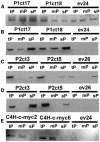
Subcellular Distribution of PAL and C4H Proteins Determined by Protein Gel Blot Analysis. Protein levels were measured in the total (tP), microsomal (mP; 130,000g pellet), and soluble (sP; 130,000g supernatant) fractions from transgenic and empty-vector control lines (15 μg protein per lane). (A) and (B) Tobacco PAL1 protein detected using anti-(tobacco PAL1) serum (A) and HA epitope-tagged tobacco PAL1 protein detected using anti-HA epitope antibody (B). P1ct17 and P1ct18 are HA-PAL1 expressing lines. (C) and (D) Tobacco PAL2 protein detected using anti-(tobacco PAL2) serum (C) and VSV-G epitope-tagged tobacco PAL2 protein identified using anti-VSV-G epitope antibody (D). P2ct3 and P2ct5 are VSV-G-PAL2–expressing lines. (E) c-myc epitope-tagged tobacco C4H protein identified using anti-c-myc epitope antibody. C4H-c-myc2 and C4H-c-myc6 are C4H-c-myc–expressing lines.
Figure 3.
Figure 3.
In Vivo Localization of PAL- and C4H-eGFP Fusion Proteins in Leaf Epidermal Cells of the Wild-Type Tobacco Plants. Transient expression of free eGFP (A), eGFP-HDEL (B), C4H-MA-eGFP (C), PAL1-eGFP (D), and PAL2-eGFP (E) was achieved by bombardment of the corresponding expression constructs. Note the reticulate pattern of localization for eGFP-HDEL, PAL1-eGFP, and C4H-MA-eGFP. Bars = 10 μm in (A) and (C) to (E) and 20 μm in (B).
Figure 4.
Figure 4.
In Vivo Localization of PAL-eGFP Fusion Proteins in Leaf Epidermal Cells of Transgenic Tobacco Plants Expressing C4H-c-myc. Transient expression of PAL1-eGFP (A) and PAL2-eGFP (B) fusion proteins in C4H-c-myc–expressing tobacco epidermal cells was achieved by bombardment of the corresponding expression constructs. Cells were also cobombarded with PAL1-eGFP and PAL2-pRTL2 (C) and PAL2-eGFP and PAL1-pRTL2 ([D] and [E]). The cell in (D) was observed 11 h after bombardment and after 15 h in (E). Note the similar reticulate pattern of localization for PAL1-eGFP in (A) and PAL2-eGFP in (B) and loss of the reticulate localization of PAL2 in (E). Bars = 20 μm in (A) to (C) and 10 μm in (D) and (E).
Figure 5.
Figure 5.
Subcellular Localization of PAL Isoforms and C4H in C4H-c-myc–Expressing Transgenic Tobacco. Protein levels were measured by gel blot analysis in the total (tP), microsomal (mP; 130,000g pellet), and soluble (sP; 130,000g supernatant) fractions (15 μg protein/lane) from C4H-cmyc–expressing and empty-vector control lines. (A) C-myc epitope-tagged tobacco C4H protein identified using anti-c-myc epitope antibody. (B) Tobacco PAL1 protein detected with anti-(tobacco PAL1) serum. (C) Tobacco PAL2 protein detected with anti-(tobacco PAL2) serum.
Figure 6.
Figure 6.
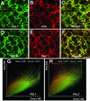
Colocalization of PAL and C4H in Leaf Protoplasts of C4H-c-myc–Expressing Tobacco. The reticulate fluorescence pattern of the Alexa Fluor-488 reporting the location of PAL1 (A) and PAL2 (D) is highly similar to the pattern of Texas Red fluorescence reporting the location of C4H ([B] and [E]). Colocalization of PAL1 with C4H (C) and PAL2 with C4H (F) is shown in the merged images. Colocalization is indicated by yellow where the green and red colors are superimposed. Arrowheads indicate typical colocalizations. The green ([A] and [D]) and red ([B] and [E]) components are depicted as two-dimensional scattergrams for PAL1-C4H (G) and PAL2-C4H (H). High colocalization coefficients were obtained for the green and red components for both interactions ([G] and [H]). Bars = 10 μm for all panels.
Figure 7.
Figure 7.
FRET Microscopy in Tobacco Protoplasts Double Labeled with Alexa Fluor-488 and Cy3. Precision FRET (pFRET) data analysis was conducted according to the methods of Elangovan et al. (2003). Single optical sections from the confocal microscope were acquired for FRET analysis using the same aperture settings, gain, and laser intensity. Double-labeled protoplasts are shown for PAL1-C4H ([A] to [D]) and PAL2-C4H ([E] to [H]). Donor excitation/donor emission and acceptor excitation/acceptor emission shows the reticulate fluorescence pattern of PAL ([A] and [E]) and C4H ([B] and [F]). Donor excitation/acceptor emission shows a fluorescence signal that includes FRET, donor cross talk, and acceptor bleed-through contaminants (uncorrected FRET) ([C] and [G]). The image after removal of donor cross talk and acceptor bleed-through represents actual FRET signal ([D] and [H]). Confocal fluorescence images of tobacco protoplasts before and after acceptor photobleaching (I). Fluorescence intensities have been pseudocolored according to the inset scale, with red pixels indicating intense fluorescence and blue pixels indicating weak fluorescence. Bars = 10 μm for all panels.

Similar articles

Cited by

References

    1. Allwood, E.G., Davies, D.R., Gerrish, C., Ellis, B.E., and Bolwell, G.P. (1999). Phosphorylation of phenylalanine ammonia-lyase: Evidence for a novel protein kinase and identification of the phosphorylated residue. FEBS Lett. 457, 47–52. - PubMed
    1. Bak, S., Kahn, R.A., Nielsen, H.L., Moller, B.L., and Halkier, B.A. (1998). Cloning of three A-type cytochromes P450, CYP71E1, CYP98, and CYP99 from Sorghum bicolor (L.) Moench by a PCR approach and identification by expression in Escherichia coli of CYP71E1 as a multifunctional cytochrome P450 in the biosynthesis of the cyanogenic glucoside dhurrin. Plant Mol. Biol. 36, 393–405. - PubMed
    1. Blount, J.W., Korth, K.L., Masoud, S.A., Rasmussen, S., Lamb, C., and Dixon, R.A. (2000). Altering expression of cinnamic acid 4-hydroxylase in transgenic plants provides evidence for a feedback loop at the entry point into the phenylpropanoid pathway. Plant Physiol. 122, 107–116. - PMC - PubMed
    1. Bolwell, G.P. (1992). A role for phosphorylation in the down-regulation of phenylalanine ammonia-lyase in suspension-cultured cells of french bean. Phytochemistry 31, 4081–4086.
    1. Bolwell, G.P., Sap, J., Cramer, C.L., Schuch, W., Lamb, C.J., and Dixon, R.A. (1985). L-Phenylalanine ammonia-lyase from Phaseolus vulgaris: Partial degradation of enzyme subunits in vitro and in vivo. Biochim. Biophys. Acta 881, 210–221.

Publication types

MeSH terms

LinkOut - more resources