Review
. 2004:20:285-308.
doi: 10.1146/annurev.cellbio.20.011403.154124.

Dorsal-ventral patterning and neural induction in Xenopus embryos

Affiliations
Review

Dorsal-ventral patterning and neural induction in Xenopus embryos

Edward M De Robertis et al. Annu Rev Cell Dev Biol. 2004.

Abstract

We review the current status of research in dorsal-ventral (D-V) patterning in vertebrates. Emphasis is placed on recent work on Xenopus, which provides a paradigm for vertebrate development based on a rich heritage of experimental embryology. D-V patterning starts much earlier than previously thought, under the influence of a dorsal nuclear -Catenin signal. At mid-blastula two signaling centers are present on the dorsal side: The prospective neuroectoderm expresses bone morphogenetic protein (BMP) antagonists, and the future dorsal endoderm secretes Nodal-related mesoderm-inducing factors. When dorsal mesoderm is formed at gastrula, a cocktail of growth factor antagonists is secreted by the Spemann organizer and further patterns the embryo. A ventral gastrula signaling center opposes the actions of the dorsal organizer, and another set of secreted antagonists is produced ventrally under the control of BMP4. The early dorsal -Catenin signal inhibits BMP expression at the transcriptional level and promotes expression of secreted BMP antagonists in the prospective central nervous system (CNS). In the absence of mesoderm, expression of Chordin and Noggin in ectoderm is required for anterior CNS formation. FGF (fibroblast growth factor) and IGF (insulin-like growth factor) signals are also potent neural inducers. Neural induction by anti-BMPs such as Chordin requires mitogen-activated protein kinase (MAPK) activation mediated by FGF and IGF. These multiple signals can be integrated at the level of Smad1. Phosphorylation by BMP receptor stimulates Smad1 transcriptional activity, whereas phosphorylation by MAPK has the opposite effect. Neural tissue is formed only at very low levels of activity of BMP-transducing Smads, which require the combination of both low BMP levels and high MAPK signals. Many of the molecular players that regulate D-V patterning via regulation of BMP signaling have been conserved between Drosophila and the vertebrates.


Figures

Figure 1
Figure 1
The Spemann-Mangold organizer experiment repeated in Xenopus laevis. (Top) control swimming tadpole; (bottom-right) Spemann organizer graft at the same stage. In the bottom-left embryo the size of the graft can be visualized as a white patch (Spemann organizer from an albino donor embryo) at early gastrula (vegetal view). The dorsal lip of the blastopore may be seen as a thin crescent opposite the graft.
Figure 2
Figure 2
Organizer formation in Xenopus laevis. At blastula stages, two signaling centers, the BCNE center in the animal region and the Nieuwkoop center in the vegetal region, pattern the embryo. Both are dependent on nuclear localization of β-Catenin on the dorsal side of the embryo. The Nieuwkoop center is formed in vegetal cells at the intersection of the VegT, Vg1, and β-Catenin gene products. The BCNE center is involved in the formation of anterior neural tissue and expresses chordin, noggin, and Xnr3. The Nieuwkoop center releases Nodal-related signals that induce Spemann's organizer in dorsal mesoderm at gastrula.
Figure 3
Figure 3
Proteins secreted by dorsal (Spemann organizer) or ventral gastrula signaling centers. Many are growth factor antagonists required for D-V patterning and are described in the text. ADMP (anti-dorsalizing morphogenetic protein) is a BMP family member that, paradoxically, is expressed in the dorsal center. Shh is sonic hedgehog and IGFBP5 is an insulin-like growth factor-binding protein that enchances IGF activity.
Figure 4
Figure 4
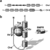
Cysteine-rich (CR) BMP-binding modules in Chordin and Crossveinless-2. (a) Chordin contains four CR modules; Crossveinless-2 (CV-2) has five. Chordin contains four additional repeats (ovals)in the region between CR1 and CR2 designated CHRD domains. These immunoglobin-like beta-barrel domains are also present in Short-gastrulation and some secreted bacterial proteins (Hyvönen 2003). CV-2 contains a von Willebrand factor-D domain (vWF-D) and a trypsin-like cysteine-rich domain (TIL) at its carboxy-terminal end. (b) A molecular complex involving BMP, Chordin, and Tsg regulates the D-V activity gradient of BMP4 in Xenopus. Chordin binds BMP4 through cysteine-rich domains CR1 and CR3. After cleavage of Chordin by the Xolloid-related (Xlr) zinc metalloprotease, the affinity between CR modules and BMP4 is greatly reduced, and BMP4 protein is able to bind to and activate BMP receptor (BMPR) on the cell surface.
Figure 5
Figure 5
Integration of multiple signaling pathways at the level of Smad1 phosphorylation during neural induction. Neural induction requires extremely low levels of Smad1 activity that are reached through the combination of two signaling systems. One is inhibition of binding between BMP4 and its serine/threonine kinase (RS/TK) receptor by anti-BMP molecules such as Chordin and Noggin. The other is inhibitory phosphorylation of Smad1 in the linker region by receptor tyrosine kinase (RTK) signals such as FGF, IGF, HGF, and EGF mediated by activation of MAPK. MH1 and MH2 are evolutionarily conserved globular Mad-homology domains; MH1 contains the DNA-binding domain and MH2 multiple protein interaction sites.
Figure 6
Figure 6
Signaling centers at blastula and gastrula that have critical roles for body plan formation in Xenopus. The BCNE center is located in the dorsal animal cap region (left) and gives rise to prospective brain and floor plate, as well as the notochord region of the Spemann organizer at gastrula (right). Nieuwkoop center cells become anterior endoderm at gastrula, coming into close apposition with prospective anterior CNS. Both signaling centers are required for brain formation. At gastrula, a ventral signaling is formed opposite the organizer.
Figure 7
Figure 7
Inversion in the course of evolution of a conserved network of extracellular regulators of BMP/Dpp signaling involved in D-V patterning.

References

    1. Agathon A, Thisse C, Thisse B. The molecular nature of the zebrafish tail organizer. Nature. 2003;424:448–52. - PubMed
    1. Agius E, Oelgeschläager M, Wessely O, Kemp C, De Robertis EM. Endodermal Nodal-related signals and mesoderm induction in Xenopus. Development. 2000;127:1173–83. - PMC - PubMed
    1. Arendt D, Nubler-Jung K. Comparison of early nerve cord development in insects and vertebrates. Development. 1999;126:2309–25. - PubMed
    1. Aubert J, Dunstan H, Chambers I, Smith A. Functional gene screening in embryonic stem cells implicates Wnt antagonism in neural differentiation. Nat. Biotechnol. 2002;20:1240–45. - PubMed
    1. Aubin J, Davy A, Soriano P. In vivo convergence of BMP and MAPK signaling pathways: impact of differential Smad1 phosphorylation on development and homeostasis. Genes Dev. 2004 In press. - PMC - PubMed

Publication types

MeSH terms

LinkOut - more resources