. 2004 Oct 8;576(1-2):174-8.
doi: 10.1016/j.febslet.2004.09.009.

Efficient assembly and release of SARS coronavirus-like particles by a heterologous expression system

Affiliations

Efficient assembly and release of SARS coronavirus-like particles by a heterologous expression system

Eduardo Mortola et al. FEBS Lett. .

Abstract

Virus-like particles (VLPs) produced by recombinant expression of the major viral structural proteins could be an attractive method for severe acute respiratory syndrome (SARS) control. In this study, using the baculovirus system, we generated recombinant viruses that expressed S, E, M and N structural proteins of SARS-CoV either individually or simultaneously. The expression level, size and authenticity of each recombinant SARS-CoV protein were determined. In addition, immunofluorescence and FACS analysis confirmed the cell surface expression of the S protein. Co-infections of insect cells with two recombinant viruses demonstrated that M and E could assemble readily to form smooth surfaced VLPs. On the other hand, simultaneous high level expression of S, E and M by a single recombinant virus allowed the very efficient assembly and release of VLPs. These data demonstrate that the VLPs are morphological mimics of virion particles. The high level expression of VLPs with correct S protein conformation by a single recombinant baculovirus offers a potential candidate vaccine for SARS.


Figures

Figure 1
Figure 1
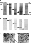
Expression of S, N, M and E SARS proteins and detection of VLPs in Sf9 infected cells. Sf9 cells were infected with single recombinant baculoviruses at a MOI of 4 and analyzed by (A) SDS–PAGE followed by Coomassie blue staining and (B) WB analysis using mouse anti‐SARS polyclonal antibody. The figure shows the clearly expressed SARS proteins when compared to the mock infected cells. It should be noted that the M protein appears as a double band in the gel and the larger form of the protein reacted poorly with our antibody. (C) EM analysis of negatively stained cell sections revealed the presence of VLPs. These particles were visible in the cytoplasm after 2 days co‐infection with the M and E expressing recombinant baculoviruses. Bar=100 nm.
Figure 2
Figure 2
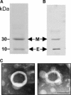
Analysis of VLPs formed by Sf9 cells co‐infected with M and E expressing recombinant baculoviruses. The VLPs were purified using a sucrose gradient and the protein composition was determined by (A) SDS–PAGE followed by Coomassie blue staining and (B) WB analysis using a mouse anti‐SARS polyclonal antibody. (C) EM of negatively stained smooth SARS VLPs purified by sucrose gradient. Bar=100 nm.
Figure 3
Figure 3
Expression of S, M and E SARS proteins in Sf9 infected cells. To examine protein expression levels, the cells were infected with the triple recombinant baculovirus at a MOI of 4 and analyzed by (A) SDS–PAGE followed by Coomassie blue staining and (B) WB analysis using mouse anti‐SARS polyclonal antibody. (C) Immunofluorescence analysis of fixed cells infected with the triple recombinant baculoviruses. Expression of the SARS S glycoprotein was detected with a primary anti‐S monoclonal antibody and secondary anti‐mouse FITC‐conjugated antibody, on the plasma membrane of infected cells. Bar=10 μm.
Figure 4
Figure 4
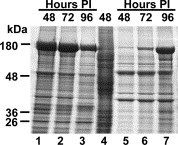
Time course of SARS‐VLPs release from Sf9 infected cells. Sf9 cells were infected with the triple recombinant baculovirus at a MOI of 4 and at the indicated times cell lysates (lanes 1–3) and cell culture medium (lanes 5–7) were analyzed by SDS–PAGE followed by Coomassie blue staining. This figure shows the location of the VLPs for purification procedures. Lane 4, Sf9 mock infected control.
Figure 5
Figure 5
Analysis of VLPs released from Sf9 cells infected with the triple recombinant baculovirus. Sucrose gradient purified VLPs from the supernatant of infected cells were analyzed by (A) SDS–PAGE followed by Coomassie blue staining and (B) WB analysis using a mouse anti‐SARS polyclonal antibody. This figure shows the protein bands of the S, M and E SARS proteins. (C) EM of the negatively stained spiky SARS‐VLPs purified by sucrose gradient from the culture medium of infected cells. Bar=100 nm. (D) Electron micrograph of immunogold‐labeled SARS‐VLPs. The VLPs were probed using an anti‐S monoclonal antibody counterstained with gold spheres coupled to anti‐mouse IgG.

References

    1. Ksiazek T.G., N. Engl. J. Med, 348, (2003), 1953– 1966. - PubMed
    1. Holmes K.V., J. Clin. Invest, 111, (2003), 1605– 1609. - PMC - PubMed
    1. Eickmann M., Science, 302, (2003), 1504– 1505. - PubMed
    1. Qin L., Acta Pharmacol. Sin, 24, (2003), 489– 496. - PubMed
    1. Ying W., Proteomics, 4, (2004), 492– 504. - PMC - PubMed

Publication types

Substances