. 2004 Nov;11(11):721-32.
doi: 10.1038/sj.cgt.7700783.

Effects of ectopic decorin in modulating intracranial glioma progression in vivo, in a rat syngeneic model

Affiliations

Effects of ectopic decorin in modulating intracranial glioma progression in vivo, in a rat syngeneic model

Alireza Biglari et al. Cancer Gene Ther. 2004 Nov.

Abstract

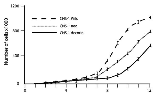
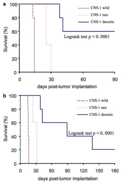
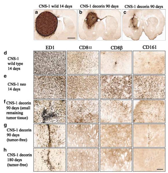
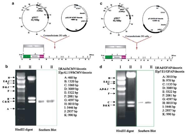
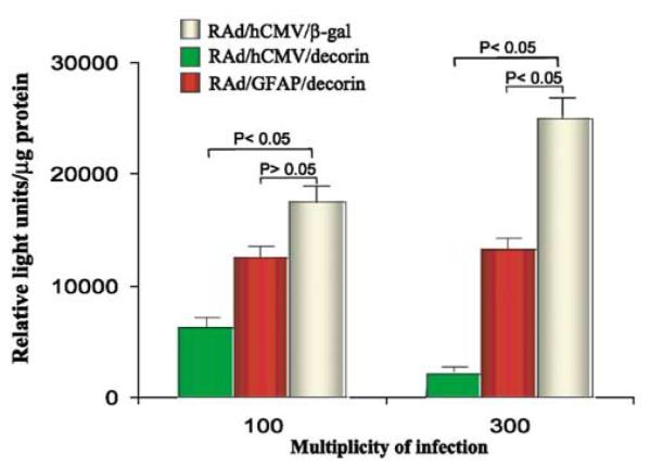
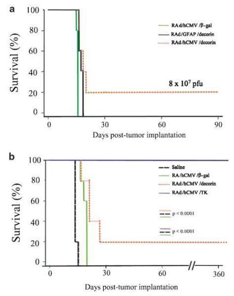
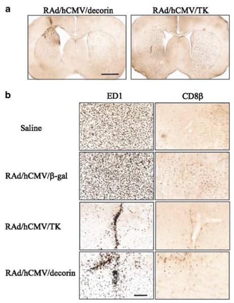
Given the failure of conventional treatments for glioblastoma, gene therapy has gained interest considerable in recent years. Gliomas are associated with a state of immunosuppression, which appears to be partially mediated by an increase in secretion of transforming growth factor-beta (TGF-beta) from glioma cells. Decorin, a small proteoglycan which can bind to and inactivate TGF-beta, has been successfully used as an antitumor strategy on stably transfected tumor cells and has been shown to cause growth suppression in neoplastic cells of various histological origins. In this paper, we investigated the use of gene therapy to deliver the decorin transgene in a site-specific manner in an experimental model of intracranial gliomas. Our aim was to inhibit the glioma-associated immunosuppressive state, and prolong the survival of tumor-bearing rats. We studied the effects of decorin gene transfer in the rat CNS-1 glioma model. To assess the effect of ectopic expression of decorin on glioma progression in vivo, stably transfected CNS-1 cells expressing decorin were implanted into the brain parenchyma of syngeneic Lewis rats. The rats implanted with CNS-1 cells expressing decorin survived significantly longer than those in the control groups which received CNS-1 cells that did not express decorin (P < .0001). We then investigated whether the survival observed with decorin expressing cells could be mimicked in vivo, using recombinant adenoviruses (RAds) expressing the decorin gene under the control of two different promoters: the human immediate-early cytomegalovirus (h-IE-CMV) and the glial fibrillary acidic protein (GFAP). In vivo results showed that administration of RAd expressing the human decorin under the control of h-IE-CMV promoter has a small, but significant effect in prolonging the survival of experimental tumor bearing rats (P < .0001). Our data indicate that ectopic decorin expression has the potential to slow glioma progression in vivo. Our results also indicate that expression of decorin has to be present in all cells which constitute the intracranial tumor mass for the inhibition of tumor growth and prolongation of the life expectancy of tumor-bearing rats to be effective.


Figures

Figure 1
Figure 1
Evaluation of bioactivity of CNS-1 cells expressing decorin. (a) Decorin expression in rat CNS-1 glioma cells. A volume of 20 μl of the PCR reaction were loaded on a 2% agarose gel containing 0.5 μg/ml ethidium bromide, and the DNA fragments fractionated by electrophoresis. The gel indicates that the decorin gene is amplified in transfected CNS-1 cells (946 bp band) compared to the negative band in control lanes; β-actin serves as an internal control; “n” is a negative control lacking genomic DNA in the PCR reaction, and “p” shows amplification from the plasmid (pAL119/decorin), used here as positive control. (b) Immunocytochemical detection of decorin in CNS-1 cells. Cells were paraformaldehyde fixed, permeabilized with Triton X-100, labeled with a sheep anti-human decorin polyclonal antibody followed by an anti-sheep FITC-conjugated secondary antibody and counterstained with 4′-6-diamidino-2 phenylindole (DAPI). The positive signal (in CNS-1 decorin cells) was not detected in neo-transfected cell samples (CNS-1 neo cells). Corresponding DAPI staining of the same cells show cell nuclei. Scale bar represents 10 μm. (c) Schematic representation of the Luciferase assay experiment. (d) Exposure of MLE/PAI/L cells to supernatant from decorin-transfected cells results in a statistically significant reduction in Luciferase activity. Results are shown as means±SEM (n=3).
Figure 2
Figure 2
In vitro proliferation of transfected CNS-1 cells. Cultured cells were subjected to daily count in quadruplicate. Growth curves were generated for each of the stably transfected and wild type CNS-1 cells over a 12-day period. Data are represented as the mean of four values (SEM is less than 10%). There was significant difference in cell-doubling times among these cell lines in vitro (P<.0001).
Figure 3
Figure 3
Kaplan–Meier survival curves for Lewis rats implanted with decorin-transfected CNS-1 cells, observed for up to 90 (a) or 180 (b) days post cell implantation of transfected or wild-type CNS-1 cells into the striatum of Lewis rats. Lewis rats implanted with CNS-1 cells expressing decorin survive significantly longer than those in the control groups (n=5, logrank test P<.0001; in both a and b).
Figure 4
Figure 4
Representative photomicrographs demonstrating brain histology and immunohistochemical staining for ED1, CD8, CD8β and CD161-positive cells in rats implanted with CNS-1 gliomas. (a) At death, all animals had large solid tumours at the site of cell implantation in the striatum. Surviving rats at 90 days post-tumor implantation had a small remaining tumor (b) or were tumor free (c). Scale bars represent 2 mm. (dh) The distribution of activated ED1 immunoreactive cells was much higher than the distribution of CD8 immunopositive cells in all rats. In the presence of a growing tumor (df), the infiltration of tumor and peritumoral tissue with inflammatory immune cells is higher in tumor-bearing animals (df) than in animals that eliminated the tumor (g,h). In animals that eliminated the tumor (g,h), only ED1+ cells remained in the visible tissue scar. Infiltration of tumors was higher in animals implanted with CNS-1 neo cells when compared to CNS-1 cells; these animals died at the same time as those implanted with control untransfected CNS-1 cells.
Figure 5
Figure 5
(a) Schematic diagram of cotransfection of pJM17 and pAL119/hCMV/decorin to generate the RAd/hCMV/decorin vector genome. (b) Southern blot analysis to verify the correct insertion of the decorin expression cassette within the RAd vector. (c) Schematic diagram of cotransfection of pJM17 and pΔE1/GFAP/decorin to generate the RAd/GFAP/decorin vector genome. (d) Southern blot analysis to confirm the correct insertion of the decorin expression cassette within the RAd vector.
Figure 6
Figure 6
Assessment of the biological activity of RAds encoding decorin. Luciferase activity in MLE/PAI/L cells following incubation with conditioned medium from HeLa cells infected with RAds encoding decorin or β-gal. Both RAd/hCMV/decorin and RAd/GFAP/decorin inhibited TGF-β signaling; RAd/hCMV/decorin was more effective than RAd/GFAP/decorin.
Figure 7
Figure 7
Kaplan–Meier survival curves for CNS-1-implanted Lewis rats treated with RAd/GFAP/decorin or RAd/hCMV/decorin. (a) At the viral dose of 8 × 10⁷, the survival rate of rats treated with RAd/hCMV/decorin was statistically better compared to rats treated with either RAd/hCMV/β-gal or RAd/GFAP/decorin (n=5, log-rank test P<.005). (b) Kaplan Meier survival curves for CNS-1 implanted Lewis rats treated with either RAd/hCMV/decorin or RAd/hCMV/HSV1-TK and ganciclovir. Lewis rats implanted with CNS-1 cells and treated with either RAd/hCMV/decorin or RAd/hCMV/HSV1-TK and ganciclovir survived significantly longer than those in control groups (n=5), with RAd/hCMV/HSV1-TK and ganciclovir being more effective than RAd/hCMV/decorin.
Figure 8
Figure 8
Representative photomicrographs demonstrating brain histology and immunohistochemical staining for ED1 and CD8β-positive cells in rat CNS-1 gliomas treated with RAd/hCMV/decorin, RAd/hCMV/HSV1-TK and ganciclovir, RAd/hCMV/β-gal or saline. (a) Enlargement of the lateral ventricle observed in an animal treated with RAd/hCMV/decorin and that survived for 12 months; this was not seen in animals that had been treated with RAd/hCMV/HSV1-TK and ganciclovir which had also survived for 12 months. (b) Rats from control groups (saline and RAd/hCMV/β-gal) had high levels of ED1+ and CD8b+ cells throughout the tumor mass, with CD8β immunoreactive cells being higher in animals injected with RAd/hCMV/β-gal. Rats that were completely tumor free at 12 months post-tumor implantation and gene therapy had infiltration of ED1-positive monocytes/macrophages in the scar remaining at the injection site, and scattered ED1 staining throughout the striatum (RAd/hCMV/HSV1-TK and RAd/hCMV/decorin). Scale bar represent 200 μm.

References

    1. Iozzo RV. Matrix proteoglycans: from molecular design to cellular function. Annu Rev Biochem. 1998;67:609–652. - PubMed
    1. Kresse H, Schonherr E. Proteoglycans of the extracellular matrix and growth control. J Cell Physiol. 2001;189:266–274. - PubMed
    1. Grant DS, Yenisey C, Rose RW, Tootell M, Santra M, Iozzo RV. Decorin suppresses tumor cell-mediated angio-genesis. Oncogene. 2002;21:4765–4777. - PubMed
    1. Yamaguchi Y, Ruoslahti E. Expression of human proteoglycan in Chinese hamster ovary cells inhibits cell proliferation. Nature. 1988;336:244–246. - PubMed
    1. Santra M, Skorski T, Calabretta B, Lattime EC, Iozzo RV. De novo decorin gene expression suppresses the malignant phenotype in human colon cancer cells. Proc Natl Acad Sci USA. 1995;92:7016–7020. - PMC - PubMed

Publication types