. 2004 Oct 12:4:39.
doi: 10.1186/1471-2148-4-39.

Evolutionary relationships of Aurora kinases: implications for model organism studies and the development of anti-cancer drugs

Affiliations

Evolutionary relationships of Aurora kinases: implications for model organism studies and the development of anti-cancer drugs

James R Brown et al. BMC Evol Biol. .

Abstract

Background: As key regulators of mitotic chromosome segregation, the Aurora family of serine/threonine kinases play an important role in cell division. Abnormalities in Aurora kinases have been strongly linked with cancer, which has lead to the recent development of new classes of anti-cancer drugs that specifically target the ATP-binding domain of these kinases. From an evolutionary perspective, the species distribution of the Aurora kinase family is complex. Mammals uniquely have three Aurora kinases, Aurora-A, Aurora-B, and Aurora-C, while for other metazoans, including the frog, fruitfly and nematode, only Aurora-A and Aurora-B kinases are known. The fungi have a single Aurora-like homolog. Based on the tacit assumption of orthology to human counterparts, model organism studies have been central to the functional characterization of Aurora kinases. However, the ortholog and paralog relationships of these kinases across various species have not been rigorously examined. Here, we present comprehensive evolutionary analyses of the Aurora kinase family.

Results: Phylogenetic trees suggest that all three vertebrate Auroras evolved from a single urochordate ancestor. Specifically, Aurora-A is an orthologous lineage in cold-blooded vertebrates and mammals, while structurally similar Aurora-B and Aurora-C evolved more recently in mammals from a duplication of an ancestral Aurora-B/C gene found in cold-blooded vertebrates. All so-called Aurora-A and Aurora-B kinases of non-chordates are ancestral to the clade of chordate Auroras and, therefore, are not strictly orthologous to vertebrate counterparts. Comparisons of human Aurora-B and Aurora-C sequences to the resolved 3D structure of human Aurora-A lends further support to the evolutionary scenario that vertebrate Aurora-B and Aurora-C are closely related paralogs. Of the 26 residues lining the ATP-binding active site, only three were variant and all were specific to Aurora-A.

Conclusions: In this study, we found that invertebrate Aurora-A and Aurora-B kinases are highly divergent protein families from their chordate counterparts. Furthermore, while the Aurora-A family is ubiquitous among all vertebrates, the Aurora-B and Aurora-C families in humans arose from a gene duplication event in mammals. These findings show the importance of understanding evolutionary relationships in the interpretation and transference of knowledge from studies of model organism systems to human cellular biology. In addition, given the important role of Aurora kinases in cancer, evolutionary analysis and comparisons of ATP-binding domains suggest a rationale for designing dual action anti-tumor drugs that inhibit both Aurora-B and Aurora-C kinases.


Figures
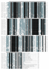
Figure 1
Figure 1
Multiple sequence alignment of representative Aurora-A (AurA), Aurora-B (AurB), and Aurora-C (AurC) kinases, and their homologs (Air1, Air2, ARK1 and Ipl1). N-terminal regions which are species-specific and could not be accurately aligned are excluded, although the numbering of residues begins at the starting amino acid for that particular peptide. Progressive darker shading indicates conservation of amino acid residues in 60%, 80% and 100% of the sequences, respectively. Dark line at the top of the sequence blocks indicates those regions used in the phylogenetic analyses (Also see additional file 1 and 2). Species include Homo sapiens (hosa), Mus musculus (mus), Danio rerio (dare), Takifugu rubripes (taru), Xenopus laevis (xela), Ciona intestinalis (ciin), Drosophila melanogaster (drme), Caenorhabditis elegans (cael), Saccharomyces cerevisiae (sace) and Schizosaccharomyces pombe (scpo). The program CLUSTALW [41] was used to constructed the initial alignment which was subsequently refined manually.
Figure 2
Figure 2
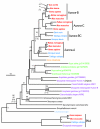
Phylogenetic tree of Aurora-A, Aurora-B, and Aurora-C kinases rooted by PLK4 kinases. Major organism groups (with colours, fonts) are mammals (red, bold italic), cold-blooded vertebrates (deep blue, italic), urochordates (orange, italic), invertebrates, (purple, italic), plants (green, italic), fungi (black, italic) and protists (light blue, italic). "Original" indicates the first Aurora identified from Drosophila melanogaster [3]. Plant sequences are identified by their Genbank accession number. Stacks of numbers show, in descending order, the percent occurrence of nodes in greater than 50% of 1000 bootstrap replicates of neighbor joining (plain text) and maximum parsimony (italicized text) analyses or greater than 50% of 10000 quartet puzzling steps of maximum likelihood analysis (in curved parentheses) or Bayesian posterior probability (only 0.90 or greater, in square parentheses). Asterisks ("*") indicate those nodes supported 70% or greater by the first three tree-building methods and 0.90 Bayesian posterior probability. Nodes with one or two values less than 50% have dashes ("-") while values less than 50% are unmarked. Scale bar represents 0.1 expected amino acid residue substitutions per site.
Figure 3
Figure 3
Comparisons of the catalytic domains of human Aurora-A, Aurora-B and Aurora-C kinases. A. Crystal structure of the catalytic domain of Human Aurora kinase with an adenosine molecule shown in the binding pocket (PDB ID 1muoA) [29]. Residues lining the active site are colored purple when invariant and red when variant. B, Multiple sequence alignment of Auroras. Using the same color scheme as the structure in panel A, residues identified to be lining the active site are identified with invariant residues among all three Auroras marked with an asterisk. Of the 26 residues lining the active site, only three vary among the different human Aurora kinases; Leu215, Thr217 and R220 (numbering and residue identity based on Aurora-A), and all of this variation was found in Aurora-A.
Figure 4
Figure 4
Unrooted phylogenetic tree of Aurora kinases from human and model organisms. Tree was constructed using the maximum likelihood quartet puzzling method [43]. Scale bar represents 0.1 expected amino acid residue substitutions per site. Confidence estimates of nodes, fonts, and colours of species names correspond to Fig. 2.

References

    1. Carmena M, Earnshaw WC. The cellular geography of aurora kinases. Nat Rev Mol Cell Biol. 2003;4:842–854. doi: 10.1038/nrm1245. - DOI - PubMed
    1. Nigg EA. Mitotic kinases as regulators of cell division and its checkpoints. Nat Rev Mol Cell Biol. 2001;2:21–32. doi: 10.1038/35048096. - DOI - PubMed
    1. Glover DM, Leibowitz MH, McLean DA, Parry H. Mutations in aurora prevent centrosome separation leading to the formation of monopolar spindles. Cell. 1995;81:95–105. doi: 10.1016/0092-8674(95)90374-7. - DOI - PubMed
    1. Reich A, Yanai A, Mesilaty-Gross S, Chen-Moses A, Wides R, Motro B. Cloning, mapping, and expression of ial, a novel Drosophila member of the Ipl1/aurora mitotic control kinase family. DNA Cell Biol. 1999;18:593–603. doi: 10.1089/104454999315141. - DOI - PubMed
    1. Schumacher JM, Golden A, Donovan PJ. AIR-2: An Aurora/Ipl1-related protein kinase associated with chromosomes and midbody microtubules is required for polar body extrusion and cytokinesis in Caenorhabditis elegans embryos. J Cell Biol. 1998;143:1635–1646. doi: 10.1083/jcb.143.6.1635. - DOI - PMC - PubMed

Publication types