. 2004 Oct 19;101(42):15184-9.
doi: 10.1073/pnas.0400751101. Epub 2004 Oct 11.

Stem cell expression of the AML1/ETO fusion protein induces a myeloproliferative disorder in mice

Affiliations

Stem cell expression of the AML1/ETO fusion protein induces a myeloproliferative disorder in mice

Timothy S Fenske et al. Proc Natl Acad Sci U S A. .

Abstract

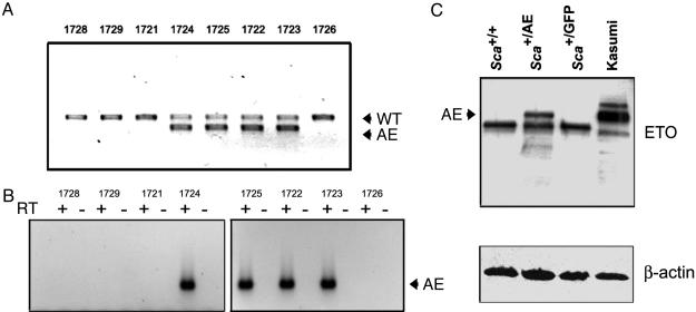
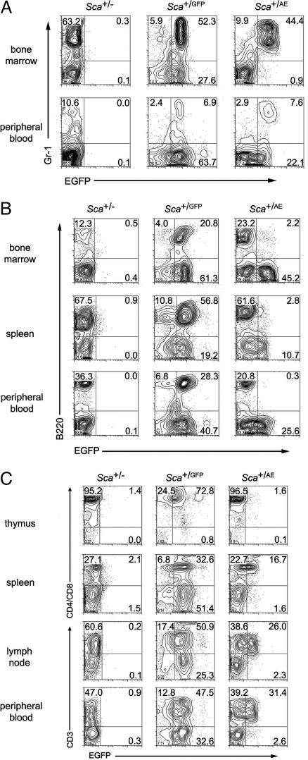
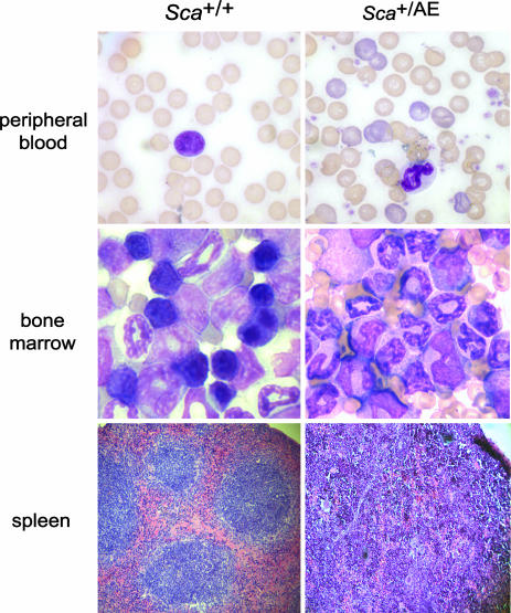
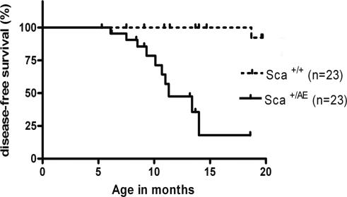
The t(8;21)(q22;q22) translocation, present in 10-15% of acute myeloid leukemia (AML) cases, generates the AML1/ETO fusion protein. To study the role of AML1/ETO in the pathogenesis of AML, we used the Ly6A locus that encodes the well characterized hematopoietic stem cell marker, Sca1, to target expression of AML1/ETO to the hematopoietic stem cell compartment in mice. Whereas germ-line expression of AML1/ETO from the AML1 promoter results in embryonic lethality, heterozygous Sca1(+/AML1-ETO ires EGFP) (abbreviated Sca(+/AE)) mutant mice are born in Mendelian ratios with no apparent abnormalities in growth or fertility. Hematopoietic cells from Sca(+/AE) mice have markedly extended survival in vitro and increasing myeloid clonogenic progenitor output over time. Sca(+/AE) mice develop a spontaneous myeloproliferative disorder with a latency of 6 months and a penetrance of 82% at 14 months. These results reinforce the notion that the phenotype of murine transgenic models of human leukemia is critically dependent on the cellular compartment targeted by the transgene. This model should provide a useful platform to analyze the effect of AML1/ETO on hematopoiesis and its potential cooperation with other mutations in the pathogenesis of leukemia.


Figures

Fig. 1.
Fig. 1.
Transgene expression. (A) Genotypes of a representative F1 litter are shown. Genomic DNA extracted from peripheral blood leukocytes was amplified in a multiplex PCR containing a shared forward primer and specific reverse primers to detect the WT or targeted (AE) alleles. WT and heterozygous Sca+/AE mice are born in Mendelian ratios. (B) AML1/ETO mRNA expression is detectable by RT-PCR in peripheral blood leukocytes from F1 heterozygous mice. The positive lanes correlate with the genotypes shown above in A. (C) Western blot analysis of bone marrow extracts from the indicated mice demonstrates expression of AML1/ETO protein in Sca+/AE mice (2.0 × 10⁵ cell equivalents per lane) and in the positive control Kasumi-1 cell line (4.0 × 10⁴ cell equivalents per lane).
Fig. 2.
Fig. 2.
Flow cytometric analysis. (A) Flow cytometric analysis was performed by using cells from mice heterozygous at the Sca1 locus for a null, GFP, or AML1/ETO ires GFP allele. Most of the myeloid (Gr1+) cells express GFP in the bone marrow and peripheral blood of Sca+/GFP and Sca+/AE mice. (B) In contrast to identically targeted Sca+/GFP mice, Sca+/AE mice demonstrate low level transgene expression in B220+ cells. (C) T cells from Sca+/AE mice express significantly lower transgene levels compared with cells from Sca+/GFP mice.
Fig. 3.
Fig. 3.
In vitro analysis. Splenocytes from Sca+/AE or WT littermate mice were cultured in RPMI medium 1640/10% FCS with IL-3 (10 ng/ml). Transgenic cells remain viable for significantly longer than WT cells. Representative data from one of three experiments are shown.
Fig. 4.
Fig. 4.
Morphologic analysis of MPD. Peripheral blood and bone marrow slides show evidence of myeloid hyperplasia with loss of erythroid and lymphoid precursors in the bone marrow and polychromasia in peripheral blood of Sca+/AE mice compared with WT littermate mice. Increased extramedullary hematopoiesis with disruption of follicular architecture is evident in the spleens of Sca+/AE mice.
Fig. 5.
Fig. 5.
Survival analysis. Kaplan-Meier plot demonstrates high penetrance and long latency of a nonlethal MPD in Sca+/AE mice compared with WT littermates (P < 0.0001).

Similar articles

Cited by

References

    1. Downing, J. R. (1999) Br. J. Haematol. 106, 296-308. - PubMed
    1. Nucifora, G. & Rowley, J. D. (1995) Blood 86, 1-14. - PubMed
    1. Okuda, T., van Deursen, J., Hiebert, S. W., Grosveld, G. & Downing, J. R. (1996) Cell 84, 321-330. - PubMed
    1. Wang, Q., Stacy, T., Binder, M., Marin-Padilla, M., Sharpe, A. H. & Speck, N. A. (1996) Proc. Natl. Acad. Sci. USA 93, 3444-3449. - PMC - PubMed
    1. Wang, Q., Stacy, T., Miller, J. D., Lewis, A. F., Gu, T. L., Huang, X., Bushweller, J. H., Bories, J. C., Alt, F. W., Ryan, G., et al. (1996) Cell 87, 697-708. - PubMed

Publication types

MeSH terms

Substances