. 2004 Nov 1;91(9):1726-34.
doi: 10.1038/sj.bjc.6602197.

Khat (Catha edulis)-induced apoptosis is inhibited by antagonists of caspase-1 and -8 in human leukaemia cells

Affiliations

Khat (Catha edulis)-induced apoptosis is inhibited by antagonists of caspase-1 and -8 in human leukaemia cells

E A O Dimba et al. Br J Cancer. .

Abstract

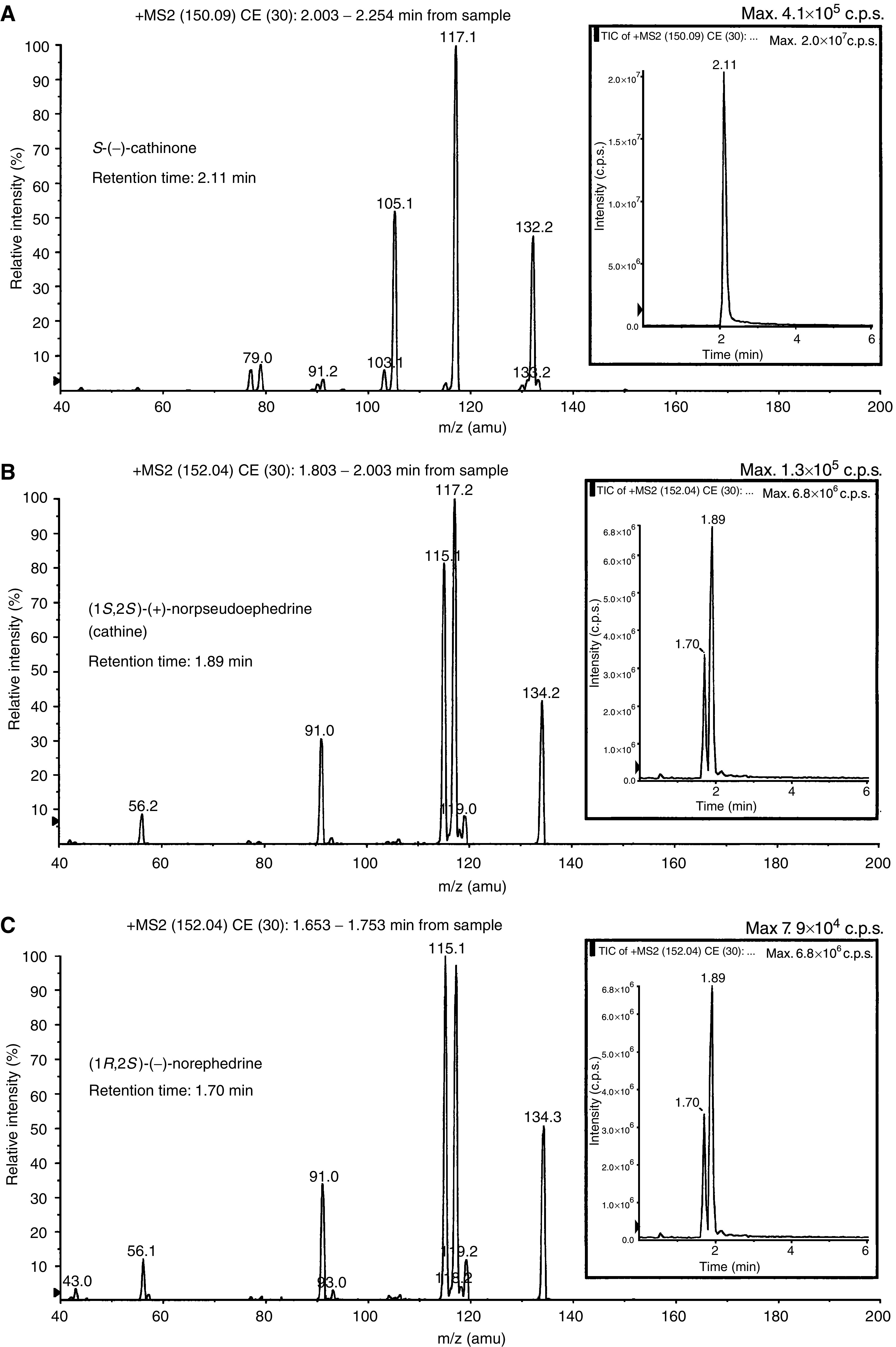
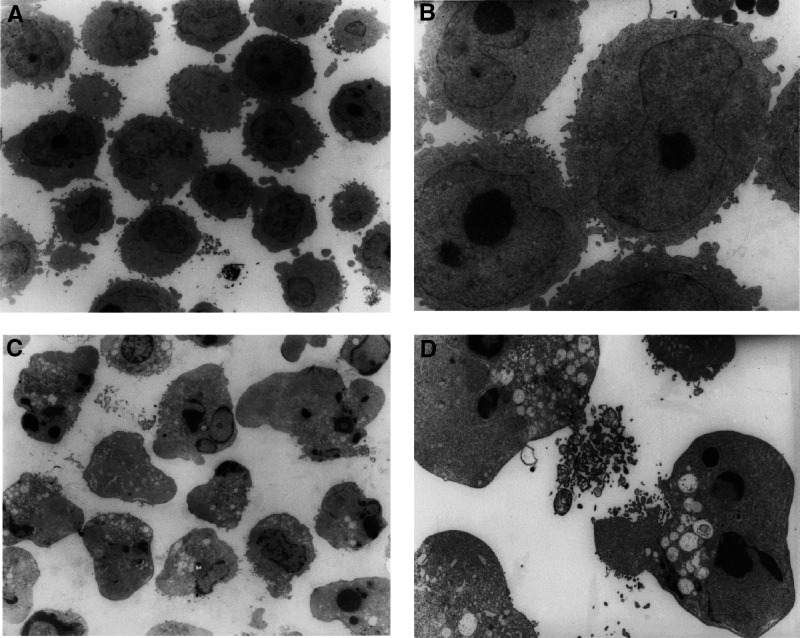
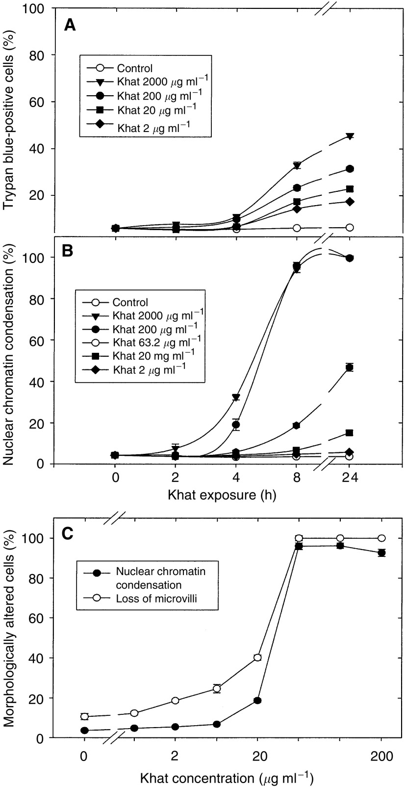
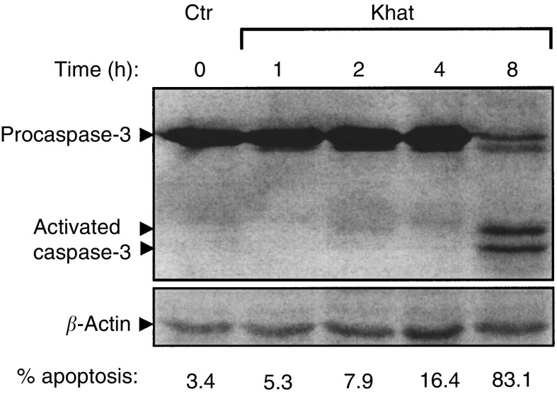
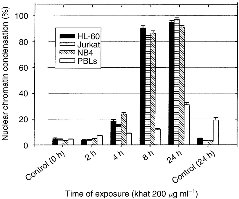
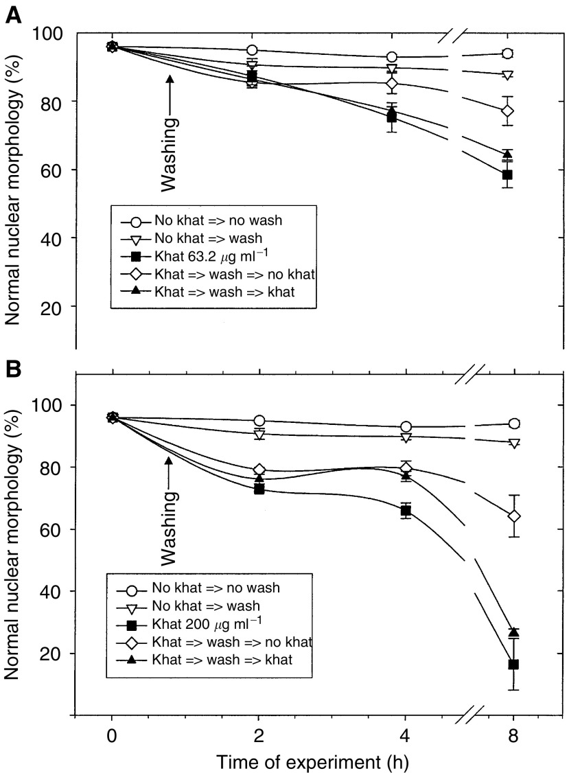
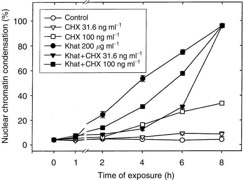
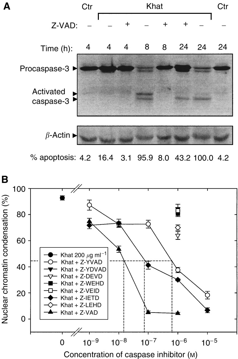
Khat chewing is a widespread habit that has a deep-rooted sociocultural tradition in Africa and the Middle East. The biological effects of khat are inadequately investigated and controversial. For the first time, we show that an organic extract of khat induces a selective type of cell death having all morphological and biochemical features of apoptotic cell death. Khat extract was shown to contain the major alkaloid compounds cathinone and cathine. The compounds alone and in combination also induced apoptosis. Khat-induced apoptosis occurred synchronously in various human cell lines (HL-60, NB4, Jurkat) within 8 h of exposure. It was partially reversed after removal of khat and the effect was dependent on de novo protein synthesis, as demonstrated by cotreatment with cycloheximide. The cell death was blocked by the pan-caspase inhibitor Z-VAD-fmk, and also by submicromolar concentrations of Z-YVAD-fmk and Z-IETD-fmk, inhibitors of caspase-1 and -8, respectively. The 50% inhibition constant (IC(50)) for khat (200 microg ml(-1))-induced apoptosis by Z-VAD-fmk, Z-YVAD-fmk and Z-IETD-fmk was 8 x 10(-7) M as compared to 2 x 10(-8) M and 8 x 10(-8) M, respectively. Western blot analysis showed a specific cleavage of procaspase-3 in apoptotic cells, which was inhibited by Z-VAD-fmk. The cell death by khat was more sensitively induced in leukaemia cell lines than in human peripheral blood leukocytes. It is concluded that khat induces a rather swift and sensitive cell death by apoptosis through mechanisms involving activation of caspase-1, -3 and -8.


Figures

Figure 1
Figure 1
LC/MS/MS of the major khat alkaloids. Mass spectrometry analysis of diluted (1 : 2000) khat extract showing specific ion scan spectra of cathinone (panel A with precursor ion m/z 150), cathine (panel B with precursor ion m/z 152) and norephedrine (panel C with precursor ion m/z 152) run at collision energy of 30 eV. Inserts: Retention time determined by total ion chromatography from the diluted khat sample.
Figure 2
Figure 2
Morphological effects of khat in HL60 cells. HL-60 cells in early logarithmic growth phase were exposed to an organic extract of khat (200 μg ml−1) for 8 h (panels B and D) or left nonsupplemented (control treated with DMSO solvent) (panels A and C). The cellular morphology was visualised by electron microscopy at × 1000 magnification (panels A–B) and at × 6000 magnification (panels C, D).
Figure 3
Figure 3
Effect of khat on cell permeability (dye exclusion test) nuclear chromatin condensation and loss of microvilli. (A, B) HL-60 cells were incubated with various concentrations of khat (range 2–2000 μg ml−1) or left nonsupplemented as controls. At various time points, cell aliquots were tested for ability to exclude trypan blue (dye exclusion test) (A) or for condensation of nuclear chromatin (B). The data represent the mean±s.e.m. of three separate experiments each in triplicate.) Cells were exposed to various concentrations (range 2–200 μg ml−1) of khat (•) or left nonsupplemented as controls (○). After exposure for 8 h, cell aliquots were fixed with formaldehyde and the fraction of cells with loss of microvilli determined. The data represent the mean±s.e.m. of three experiments in triplicate.
Figure 4
Figure 4
Activation of procaspase-3 in khat-exposed cells. Cell lysates were prepared and proteins separated by SDS–PAGE and transblotted onto PVDF membranes. Caspase-3 was probed by mouse α-caspase-3 and detected using a horseradish peroxidase-coupled secondary antibody (see Materials and Methods). β-Actin was used as an internal reference marker to monitor the amount of protein in each lane.
Figure 5
Figure 5
Human leukaemia cell lines undergo cell death (apoptosis) more sensitively than peripheral blood human leukocytes (PBLs) after exposure to khat. Various human acute myeloid (HL-60, NB4) and lymphoblastic leukaemic (Jurkat) cell lines as well as isolated PBLs were exposed to 200 μg ml−1 khat for various time points or left nonsupplemented as controls. The fraction of cells with condensed nuclear chromatin was determined. The data on the cell lines represent the mean±s.e.m. of one experiment in triplicates, whereas the data on PBLs were obtained from three healthy individuals, each run in triplicates. The differences between khat-treated and untreated cells, and between khat-treated cell lines and PBLs were statistically significant (P<0.05).
Figure 6
Figure 6
Test on commitment to cell death after short-term exposure to khat. HL-60 cells were exposed to 63.2 μg ml−1 khat (A) or 200 μg ml−1 khat (B) for 0.5 h, washed, and left nonsupplemented (open symbols) or resupplemented (filled symbols) with khat. Some cultures were treated continuously with 63.2 μg ml−1 khat (▪, panel A) or 200 μg ml−1 khat (▪, panel B) as reference. Other cultures were left unsupplemented with khat with (▿) or without washing (○). At the times indicated, cell aliquots (50 μl) were mixed with one volume fixative. Cell survival (%) was based on determination of the fraction of cells not having condensed nuclear features. These morphological findings were considered typical of cells irreversibly committed to death. The data represent the mean±s.e.m. of triplicate experiments. The difference between khat-treated samples and untreated (controls) were statistically significant (P<0.05).
Figure 7
Figure 7
Khat-induced cell death in HL-60 cells was dependent on de novo protein synthesis. Cells were exposed to 200 μg ml−1 khat in the absence (•) or presence of 31.6 ng ml−1 CHX (▾) or 100 ng ml−1 CHX (▪). Some cultures were left unsupplemented (○) as controls or treated with 31.6 ng ml−1 CHX (▿) or 100 ng ml−1 CHX (□). CHX was added 15 min prior to treatment with khat. At the times indicated, cell aliquots (50 μl) were removed and dead cells determined as the fraction of cells (%) having condensed nuclear chromatin. The data represent means±s.e.m. of two separate experiments, each in triplicate.
Figure 8
Figure 8
Selective inhibition of khat-induced cell death by caspase inhibitors. (A) Cultures of HL-60 (10 × 106) cells were exposed with 200 μg ml−1 khat for various time points in the absence or presence of 10−6M Z-VAD-fmk or left nonsupplemented as control cultures. (B) HL-60 cells were treated with 200 μg ml−1 khat for 8 h in the absence () or presence of various concentrations of Z-VAD-fmk (), a pan-caspase inhibitor, or the caspase selective inhibitors Z-YVAD-fmk () and Z-IETD-fmk (), inhibiting caspase-1 and -8, respectively. For the caspase inhibitors Z-VDVAD-fmk, Z-DEVD-fmk, Z-WEHD-fmk, Z-VEID-fmk and Z-LEHD-fmk, selecting predominantly inhibiting caspases-2, -3, -5, -6 and -9, respectively, only the effect of 10−6M concentration of these inhibitors is shown. Half maximal inhibitory concentration (ID50) for induction of cell death by 200 μg ml−1 khat extract after 8 h of exposure was determined to be 2 × 10⁻⁸, 9 × 10⁻⁸ and 8 × 10⁻⁷M for Z-VAD, Z-IETD and Z-YVAD, respectively. The data represent means±s.e.m. of two separate experiments, each in triplicates.

References

    1. Al-Ahdal MN, McGarry TJ, Hannan M (1988) Cytoxicity of khat (Catha edulis) extract on cultured mammalian cells: effects on macromolecule biosynthesis. Mutat Res 204: 317–322 - PubMed
    1. Al-Mamary M, Al-Habori M, Al-Aghbari AM, Baker MM (2002) Investigation into the toxicological effects of Catha edulis leaves: a short term study in animals. Phytother Res 16: 127–132 - PubMed
    1. Al-Meshal I, Qureshi S, Ageel AM, Tariq M (1991) The toxicity of Catha edulis in mice. J Subst Abuse 3: 107–115 - PubMed
    1. Al-Motarreb A, Baker K, Broadley KJ (2002) Khat: pharmacological and medical aspects and its social use in Yemen. Phytother Res 16: 403–413 - PubMed
    1. Al-Qirim TM, Shahwan M, Zaidi KR, Uddin Q, Banu N (2002) Effect of khat, its constituents and restraint stress on free radical metabolism of rats. J Ethnopharmacol 83: 245–250 - PubMed

Publication types

MeSH terms