. 2005 Apr 1;387(Pt 1):129-38.
doi: 10.1042/BJ20041359.

Prelamin A endoproteolytic processing in vitro by recombinant Zmpste24

Affiliations

Prelamin A endoproteolytic processing in vitro by recombinant Zmpste24

Douglas P Corrigan et al. Biochem J. .

Abstract

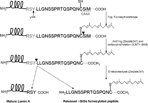
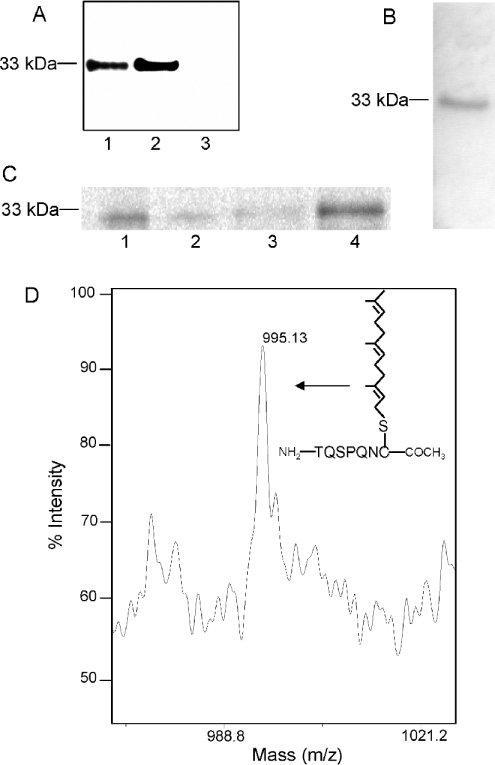
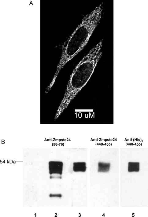
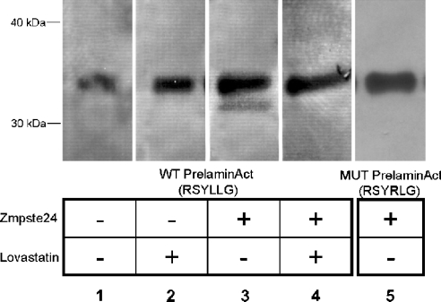
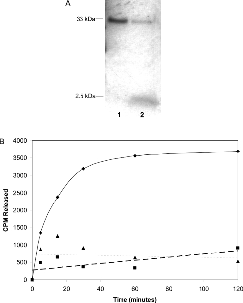
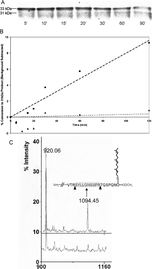
The nuclear lamins form a karyoskeleton providing structural rigidity to the nucleus. One member of the lamin family, lamin A, is first synthesized as a 74 kDa precursor, prelamin A. After the endopeptidase and methylation reactions which occur after farnesylation of the CAAX-box cysteine, there is a second endoproteolysis that occurs 15 amino acids upstream from the C-terminal farnesylated cysteine residue. Studies with knockout mice have implicated the enzyme Zmpste24 (Face-1) as a suitable candidate to perform one or both of these proteolytic reactions. Evidence has been presented elsewhere establishing that Zmpste24 possesses a zinc-dependent CAAX endopeptidase activity. In the present study, we confirm this CAAX endopeptidase activity with recombinant, membrane-reconstituted Zmpste24 and show that it can accept a prelamin A farnesylated tetrapeptide as substrate. To monitor the second upstream endoproteolytic cleavage of prelamin A, we expressed a 33 kDa prelamin A C-terminal tail in insect cells. We demonstrate that this purified substrate possesses a C-terminal farnesylated and carboxyl-methylated cysteine and, therefore, constitutes a valid substrate for assaying the second endoproteolytic step in lamin A maturation. With this substrate, we demonstrate that insect cell membranes bearing recombinant Zmpste24 can also catalyse the second upstream endoproteolytic cleavage.


Figures

Figure 1
Figure 1. The processing pathway of prelamin A
Prelamin A undergoes a series of post-translational modifications at the designated CAAX box, CSIM, including farnesylation, AAX endopeptidolysis, carboxyl-methylation and a second endoproteolytic cleavage between a tyrosine and a leucine residue, 15 amino acids upstream from the farnesylated cysteine residue. This results in the production of mature lamin A, along with an approx. 2 kDa released prenylated peptide.
Figure 2
Figure 2. CAAX box modification of membrane-associated prelaminAct expressed in insect cells
PrelaminAct fused to His6 at the N-terminus was cloned and expressed in insect cells by means of baculovirus. The expressed prelaminAct is solubilized from membranes and purified by binding and elution from Ni-agarose beads as described in the Experimental section. (A) SDS/PAGE and immunoblotting with anti-prelamin A of purified prelaminAct cytosolic fraction (lane 1), membrane fraction (lane 2) and mock-transfected purification (lane 3). (B) Metabolic labelling of prelaminAct with [3H]mevalonate. Prelamin A was isolated by SDS/PAGE and visualized by fluorography. (C) Immunoprecipitation with anti-farnesyl antibody of purified cytosolic and membrane-associated prelaminAct metabolically labelled with [35S]methionine. Immunoprecipitates (lanes 1 and 3) and supernatants (lanes 2 and 4) of membrane (lanes 1 and 2) and cytosolic fractions (lanes 3 and 4) were analysed by SDS/PAGE and visualized by fluorography. (D) MALDI–TOF analysis of Ni-agarose- and SDS/PAGE-purified membrane-associated prelaminAct. The 33 kDa band was excised from the gel and digested with trypsin. The C-terminal peptide (depicted in the inset) modified by AAXing, farnesylation and carboxyl-methylation (molecular mass, 995.13 Da) was observed.
Figure 3
Figure 3. Cloning and expression of Zmpste24
(A) Subcellular localization of Zmpste24 expressed in mammalian cells. Zmpste24 was cloned from a HeLa cDNA library into the mammalian expression vector pcDNA3.1 and transiently transfected into CHO-K1 cells. Subcellular localization was determined by indirect immunofluorescence with anti-Zmpste24 (residues 56–76). Note both the cytosolic and perinuclear distribution of Zmpste24. (B) Immunoblot with anti-Zmpste24 (residues 56–76; lanes 1–3), anti-Zmpste24 (residues 440–455; lane 4) or anti-His6 (lane 5) of expressed and Ni-agarose-purified Zmpste24 from insect cells. Lane 1, Ni-agarose-purified elution of mock-transfected cells; lane 2, total cell extract before purification; lanes 3–5, Ni-agarose-purified elutions of Zmpste24.
Figure 4
Figure 4. Zmpste24 processing of prelaminAct in transfected insect cells
Zmpste24 was virally transfected into insect cells expressing N-terminally His6-tagged prelaminAct or prelaminAct mutated at the endoproteolytic cleavage site (L647R). The cells were then incubated for 72 h in the presence or absence of lovastatin. PrelaminAct and laminAct were purified using Ni-agarose chromatography, separated by SDS/PAGE and visualized by immunoblotting with anti-lamin A.
Figure 5
Figure 5. Zmpste24-catalysed cleavage of a 2 kDa fragment from prelaminAct
(A) PrelaminAct was metabolically labelled for 16 h with [3H]mevalonate, 48 h after infecting with the prelaminAct construct. PrelaminAct was then purified with Ni-agarose. Products formed after incubation with either mock-transfected (lane 1) or Zmpste24 (lane 2) membranes were separated by SDS/PAGE and visualized by fluorography. (B) Purified prelaminAct from insect cell cytosol was in vitro farnesylated with [3H]FPP and farnesyltransferase and immobilized on Ni-agarose beads. The beads were incubated with membranes from Zmpste24 or mock-transfected cells or no enzyme. At various times, aliquots of the supernatant were counted to determine release of the farnesylated C-terminus of prelamin A (◆, Zmpste24 membranes; ■, mock-infected; ▲, no membranes).
Figure 6
Figure 6. Zmpste24 can endoproteolytically process prelaminAct at the sequence RSYLLG
PrelaminAct or its mutant (L647R) were metabolically labelled with [35S]methionine in insect cells and purified with Ni-agarose. The proteins were incubated with either Zmpste24 or mock-transfected membranes in the presence of E-64 for various times. (A) WT (wild-type) prelaminAct substrate and endoproteolytic products were separated by SDS/PAGE and visualized by phosphoimager analysis. (B) Relative formation of the 31 kDa product ([31 kDa counts]/[33 kDa+31 kDa counts]–background) as a function in time. Background is defined as the counts observed for mock-transfected membranes incubated with WT substrate. Products continue to be formed for at least 2 h (■, Zmpste24 membranes with WT prelaminAct; ▲, Zmpste24 membranes with RSYRLG mutant prelaminAct). (C) MALDI–TOF analysis of tryptic digest of 33 kDa (upper spectrum) and 31 kDa (lower spectra) gel excised bands, indicating the disappearance of the peak corresponding to the peptide SYLLGNSSPR (molecular mass 1094.21 Da). The peak appearing at 920.06 Da corresponds to the predicted peptide from the prelaminAct sequence between residues 428 and 435. The inset depicts the predicted C-terminal sequence of prelaminAct along with the appropriate CAAX modifications. Arrows represent tryptic cleavage sites. The released peptide (SYLLGNSSPR) has a predicted molecular mass of 1094.45 Da.

References

    1. Glomset J. A., Gelb M. H., Farnsworth C. C. Prenyl proteins in eukaryotic cells: a new type of membrane anchor. Trends Biochem. Sci. 1990;15:139–142. - PubMed
    1. Rando R. R. Chemical biology of protein isoprenylation/methylation. Biochim. Biophys. Acta. 1996;1300:5–16. - PubMed
    1. Zhang F. L., Casey P. J. Protein prenylation: molecular mechanisms and functional consequences. Annu. Rev. Biochem. 1996;65:241–269. - PubMed
    1. Sinensky M. Recent advances in the study of prenylated proteins. Biochim. Biophys. Acta. 2000;1484:93–106. - PubMed
    1. Beck L. A., Hosick T. J., Sinensky M. Isoprenylation is required for the processing of the lamin A precursor. J. Cell Biol. 1990;110:1489–1499. - PMC - PubMed

Publication types

MeSH terms

LinkOut - more resources