. 2004 Nov;53(11):1632-8.
doi: 10.1136/gut.2003.037911.

Induction of T lymphocyte apoptosis by sulphasalazine in patients with Crohn's disease

Affiliations

Induction of T lymphocyte apoptosis by sulphasalazine in patients with Crohn's disease

J Doering et al. Gut. 2004 Nov.

Abstract

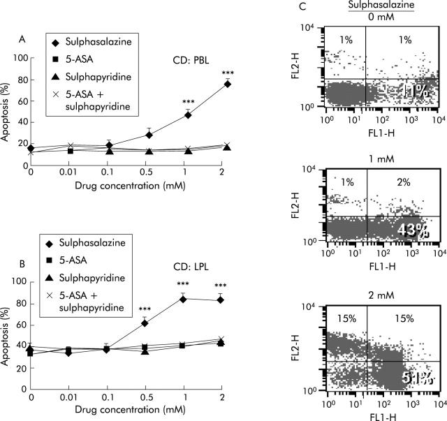
Background: Lamina propria T lymphocytes (LPL) of the intestinal mucosa are chronically activated in Crohn's disease (CD). Defective apoptosis of activated LPL was proposed as a key pathogenic mechanism. In fact, increased expression of antiapoptotic molecules was observed in CD LPL. In the present work, we aimed to analyse the effects and underlying molecular mechanisms of 5-amino salicylic acid (5-ASA) and derivatives on apoptosis of LPL and peripheral blood lymphocytes (PBL) in patients with CD compared with ulcerative colitis (UC) and in non-inflammatory controls.

Methods: PBL and LPL were isolated by Ficoll-Hypopaque gradient centrifugation and the EGTA-collagenase method, respectively. PBL/LPL were stimulated with FasL, 5-ASA, sulphapyridine, and sulphasalazine for 24/48 hours and apoptosis was quantified by flow cytometry (annexin V- propidium iodide method) and immunofluorescence. The molecular mechanisms of drug induced apoptosis were analysed in wild-type and FADD-/- Jurkat T cells using western blots and caspase assays.

Results: While PBL displayed a normal apoptosis pattern after Fas stimulation in patients with active CD, LPL from inflammatory areas were highly resistant. Comparable resistance to apoptosis was observed in LPL of UC patients. In contrast with 5-ASA, which did not induce apoptosis in lymphocytes, sulphasalazine proved to be a potent proapoptotic agent. Sulphasalazine induced T lymphocyte apoptosis was independent of the Fas pathway but associated with marked downregulation of antiapoptotic bcl-xl and bcl2, activation of the mitochondrial apoptosis signalling pathway, and subsequent activation of caspase-9 and caspase-3.

Conclusion: The beneficial effect of sulphasalazine in treating inflammatory bowel disease is at least in part attributable to its proapoptotic effects on LPL which allows potent downregulation of lymphocyte activation.


Figures

Figure 1
Figure 1
Response to Fas stimulation (FasLigand (FasL)) in lamina propria T lymphocytes (LPL) and peripheral blood lymphocytes (PBL). (A) LPL derived from inflammatory lesions of Crohn’s disease (CD) and ulcerative colitis (UC) patients but not LPL from non-inflammatory controls showed a marked resistance to Fas induced apoptosis. In contrast (B), PBL from CD as well as from UC displayed a high degree of apoptosis after Fas stimulation comparable with controls. (C) Comparison of LPL derived from inflammatory and non-inflammatory sections in three CD patients: LPL of inflammatory areas were resistant to Fas induced apoptosis. Similarly, after pre-stimulation with interleukin 2 (IL-2 50 IU/ml for seven days), cytokine withdrawal induced only apoptosis in LPL from non-inflammatory sections whereas LPL from inflamed tissue were resistant to IL-2 withdrawal induced apoptosis. (D) Time-kinetic studies in Fas stimulated PBL revealed an identical apoptosis pattern for inflammatory CD and control patients.
Figure 2
Figure 2
Effect of sulphasalazine on peripheral blood lymphocyte (PBL)/lamina propria T lymphocyte (LPL) apoptosis. Stimulation with sulphasalazine induced dose dependent apoptosis in (A) PBL or (B) LPL from inflammatory lesions in Crohn’s disease (CD) patients. In contrast, sulphapyridine, 5-amino salicylic acid (5-ASA) or the combination of both drugs had no proapoptotic effect at any concentration tested. (C) Flow cytometric analysis of PBL from an inflammatory CD patient allowed quantification of early and late apoptotic events after sulphasalazine stimulation using the annexin V-propidium iodide assay (see material and methods). ***p<0.001.
Figure 3
Figure 3
Role of Fas/Fas associated death domain protein (FADD) in sulphasalazine induced apoptosis. (A) Jurkat T cells responded with apoptosis after sulphasalazine stimulation in a way similar to peripheral blood lymphocytes (PBL)/lamina propria T lymphocytes (LPL). Wild-type (WT) and FADD-neg Jurkat showed the same apoptotic response to sulphasalazine. In contrast, 5-amino salicylic acid (5-ASA), sulphapyridine, 4-ASA, or NAC failed to induce Jurkat apoptosis (B). (C) Morphological analysis of sulphasalazine induced apoptosis (immunofluorescence after HOECHST33342 staining) revealed nuclear condensation and fragmentation of treated cells comparable with Fas induced apoptosis. (D) FADD-neg Jurkat cells were resistant to Fas induced apoptosis confirming that sulphasalazine induced apoptosis was independent of a functional death inducing signalling complex.
Figure 4
Figure 4
Mechanisms of sulphasalazine induced apoptosis. (A) Sulphasalzine induced a shift in bcl homeostasis towards a proapoptotic constellation by reducing antiapoptotic bcl-xl and bcl 2 expression, without affecting expression of proapoptotic bax or bak, as shown by western blot analysis. In contrast, 5-amino salicylic acid (5-ASA), sulphapyridine, alone or in combination, did no alter any bcl molecule expression. (B) Activation of caspase-9 in response to sulphasalazine was demonstrated in an indirect way by disappearance of the proenzyme at 48/46 kDa; 5-ASA and sulphapyridine had no effect. (C) Inhibition of the caspase cascade using the broad range caspase inhibitor zVAD-fmk blocked sulphasalazine induced apoptosis, as did the caspase-3 specific inhibitor zDEVD-fmk. (D) Activation of caspase-3 after sulphasalazine stimulation was monitored by western blot, showing the occurrence of the active enzymes at 17 and 12 kDa with the proenzyme migrating at 32 kDA. Again, 5-ASA, sulphapyridine, and the combination of both drugs had no effect on caspase-3. One of three representative experiments is shown.

Comment in

References

    1. Fiocchi C . Inflammatory bowel disease: etiology and pathogenesis. Gastroenterology 1998;115:182–205. - PubMed
    1. Podolsky DK. Inflammatory bowel disease. N Engl J Med 2002;347:417–29. - PubMed
    1. Shanahan F . Crohn’s disease. Lancet 2002;359:62–9. - PubMed
    1. Levine AD. Apoptosis: Implications for inflammatory bowel disease. IBD 2000;6:191–205. - PubMed
    1. Ina K , Itoh J, Fukushima K, et al. Resistance of Crohn’s disease T cells to multiple apoptotic signals is associated with a Bcl-2/Bax mucosal imbalance. J Immunol 1999;163:1081–90. - PubMed

Publication types

MeSH terms