. 2004 Nov;53(11):1688-97.
doi: 10.1136/gut.2003.031997.

Antidiabetic thiazolidinediones inhibit invasiveness of pancreatic cancer cells via PPARgamma independent mechanisms

Affiliations

Antidiabetic thiazolidinediones inhibit invasiveness of pancreatic cancer cells via PPARgamma independent mechanisms

A Galli et al. Gut. 2004 Nov.

Abstract

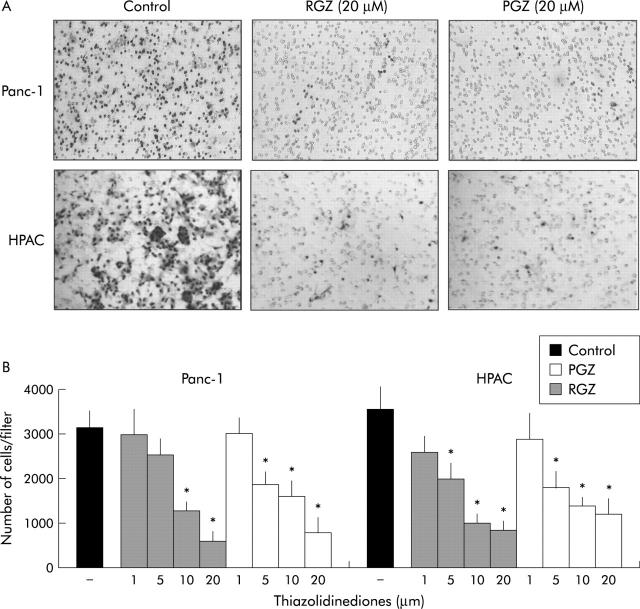
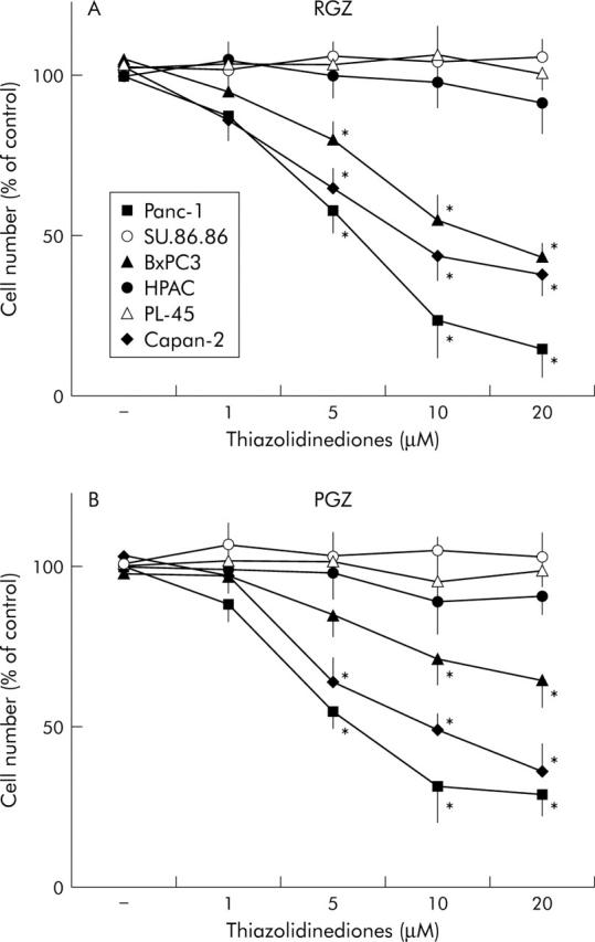
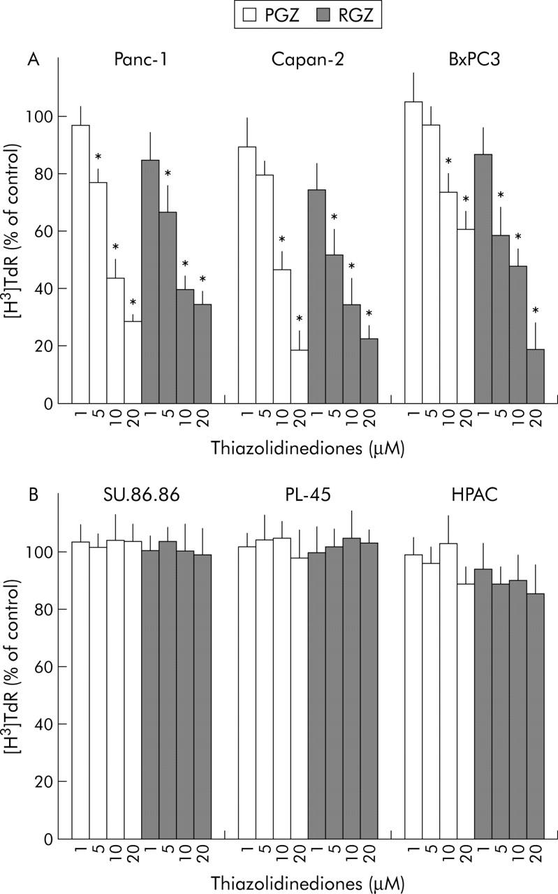
Background/aims: Thiazolidinediones (TZD) are a new class of oral antidiabetic drugs that have been shown to inhibit growth of some epithelial cancer cells. Although TZD were found to be ligands for peroxisome proliferators activated receptor gamma (PPARgamma) the mechanism by which TZD exert their anticancer effect is currently unclear. Furthermore, the effect of TZD on local motility and metastatic potential of cancer cells is unknown. The authors analysed the effects of two TZD, rosiglitazone and pioglitazone, on invasiveness of human pancreatic carcinoma cell lines in order to evaluate the potential therapeutic use of these drugs in pancreatic adenocarcinoma.

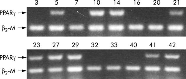
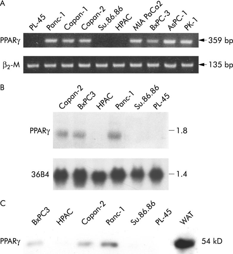
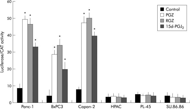
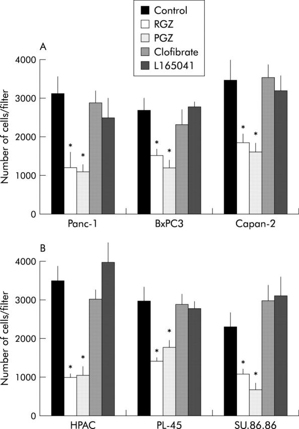
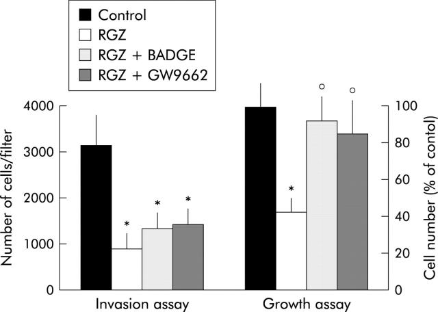
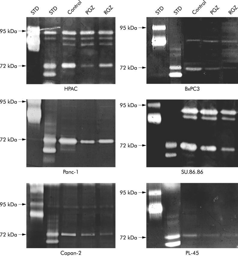
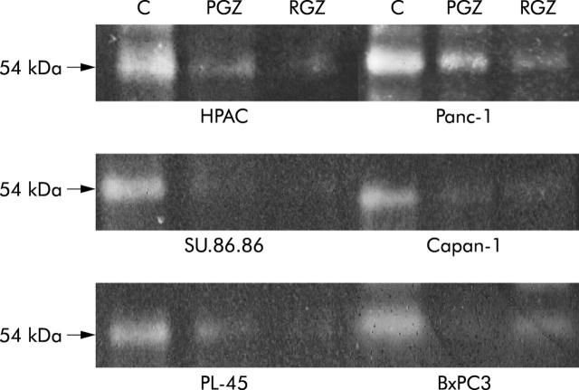
Methods: Expression of PPARgamma in human pancreatic adenocarcinomas and pancreatic carcinoma cell lines was measured by reverse transcription polymerase chain reaction and confirmed by western blot analysis. PPARgamma activity was evaluated by transient reporter gene assay. Invasion assay was performed in modified Boyden chambers. Gelatinolytic and fibrinolytic activity were evaluated by gel zymography.

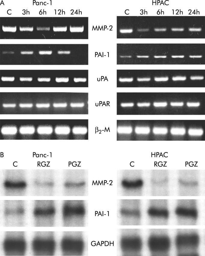
Results: TZD inhibited pancreatic cancer cells' invasiveness, affecting gelatinolytic and fibrinolytic activity with a mechanism independent of PPARgamma activation and involving MMP-2 and PAI-1 expression.

Conclusion: TZD treatment in pancreatic cancer cells has potent inhibitory effects on growth and invasiveness suggesting that these drugs may have application for prevention and treatment of pancreatic cancer in humans.


Figures

Figure 1
Figure 1
PPARγ expression in human pancreatic adenocarcinoma. One microgram of total RNA extracted from 16 representative human pancreatic adenocarcinomas was reverse transcribed using random hexamers and amplified by polymerase chain reaction using specific primers for PPARγ and for β2-microglobulin (β2-M) as described in Methods. The reverse transcription polymerase chain reaction products were electrophoresed on ethidium bromide containing agarose gel. Numbers correspond to patient number in table 1 ▶.
Figure 2
Figure 2
PPARγ expression in cultured human pancreatic adenocarcinoma cells. (A) RT-PCR analysis of PPARγ in 10 pancreatic cancer cell lines. One microgram of total RNA was reverse transcribed using random hexamers and amplified by polymerase chain reaction using specific primers for PPARγ and β2-microglobulin (β2-M). The reverse transcription polymerase chain reaction products were electrophoresed on ethidium bromide containing agarose gel. (B) Northern blot analysis of total RNA (20 μg) extracted from three PPARγ expressing (Panc-1, BxPC-3, Capan-2) and three non-expressing (HPAC, SU.86.86, PL-45) cell lines. Denaturated RNA sample were electophoresed in 1% agarose-3% formaldehyde gel and transferred to a nylon membrane. Filters were prehybridised and then hybridised with the complementary PPARγ and 36B4 cDNA probes as described in Methods. (C) Levels of immunoreactive PPARγ in pancreatic cancer cells. Fifty micrograms of nuclear protein extracts were fractionated by sodium dodecyl sulfate-polyaclylamide gel electrophoresis and transferred to nitrocellulose paper. Receptor proteins were detected by incubating the filter with specific anti-PPARγ antibody. Total protein extracts from human white adipose tissue (WAT) was used as positive control.
Figure 3
Figure 3
PPARγ transcriptional activity in human pancreatic adenocarcinoma cells. After overnight attachment, cells were transfected with ARE-73-tk-luciferase reporter plasmid and pSV2-CAT as internal control for transfection efficiency. Twenty four hours after transfection cells were treated with 20 μM of TZD (RGZ or PGZ) or 5 μM of 15d-PGJ2 or vehicle (control). Twenty four hours after treatment the cells were harvested for luciferase and CAT assay as described in Methods. The data are expressed as mean (SD) for three or four replicate experiments performed in triplicate; *denotes statistical significance (p<0.01 or higher degree of significance) versus control.
Figure 4
Figure 4
Thiazolidinediones inhibit the invasiveness of cultured pancreatic cancer cells independently of PPARγ. Confluent cells were serum starved for 24 hours and then exposed to the different receptor ligands or vehicle for one hour. Cells were then trypsinised, resuspended in serum free medium with or without ligands (20 μM TZD or 1 mM clofibric acid or 1 μM L165041), and placed in the upper compartments of Boyden chambers. After six hours’ incubation polycarbonate filters were fixed in methanol and stained with haematoxylin and eosin. Cells were then counted with a computerised video image system. (A) PPARγ expressing cell lines. (B) PPARγ non-expressing cell lines. The mean (SD) of six independent experiments performed for each in duplicate are shown; *denotes statistical significance (p<0.03 or higher degree of significance) versus vehicle treated controls.
Figure 5
Figure 5
Thiazolidinediones inhibit the invasiveness of cultured pancreatic cancer cells in a dose dependent manner. Confluent cells were serum starved for 24 hours and then exposed to TZD or vehicle (RGZ or TGZ) for one hour. Cells were then trypsinised, resuspended in serum free medium with or without TZD at the concentration indicated, and placed in the upper compartments of Boyden chambers. After six hours’ incubation polycarbonate filters were fixed in methanol and stained with haematoxylin and eosin. Cells were then counted with a computerised video image system. (A) A representative picture showing inhibition of invading cells after treatment with 20 μM of TZD. Original magnification ×200. (B) TZD treatments inhibit the invasiveness of pancreatic cancer cells in a dose dependent manner. The mean (SD) of six independent experiments performed for each in duplicate are shown; *denotes statistical significance (p<0.05 or higher degree of significance) versus vehicle treated controls.
Figure 6
Figure 6
Effect of TZD treatment on anchorage dependent growth in human pancreatic adenocarcinoma cells. Cells were counted after 72 hours of incubation with TZD at appropriate concentrations. Control cells received vehicle alone. After trypsinisation cells were resuspended in phosphate buffer saline and counted in a hemocytomer as described in Methods. (A) Treatment with rosiglitazone (RGZ). (B) Treatment with pioglitazone (PGZ). PPARγ non-expressing cell lines. Cell number was expressed as percentage of untreated controls. The mean (SD) of six independent experiments each performed in duplicate or triplicate are shown; *denotes statistical significance (p<0.05 or higher degree of significance) versus control.
Figure 7
Figure 7
Effect of TZD treatment on DNA synthesis in human pancreatic adenocarcinoma cells. [3H]TdR incorporation: subconfluent cells were incubated in serum free/insulin free medium for 24 hours and then treated with increased concentrations of TZD (RGZ or PGZ) or their vehicle for 42 hours. After a six hour pulsing period with [3H]TdR (1.0 μCi/mL; see Methods for details) the cells were harvested. (A) PPARγ expressing cell lines. (B) PPARγ non-expressing cell lines. Data were expressed as percentage of untreated controls. The mean (SD) of six independent experiments each performed in triplicate are shown; *denotes statistical significance (p<0.05 or higher degree of significance) versus control.
Figure 8
Figure 8
Effects of PPARγ antagonists on growth and invasiveness of Panc-1 cells. For the invasion assay confluent Panc-1 cells were serum starved for 24 hours and then exposed to 20 μM RGZ or vehicle with or without PPARγ antagonist (20 μM BADGE or 10 μM GW9662) for one hour. Cells were then trypsinised, resuspended in serum free medium with or without RGZ and PPARγ antagonists, and placed in the upper compartments of Boyden chambers. After six hours’ incubation polycarbonate filters were fixed in methanol and stained with haematoxylin and eosin. Cells were then counted with a computerised video image system. Cells growth was evaluated by cell counting after 72 hours of incubation with 20 μM RGZ with or without PPARγ antagonists (20 μM BADGE or 10 μM GW9662). Control cells received vehicle alone. After trypsinisation cells were resuspended in phosphate buffer saline and counted in a hemocytomer as described in Methods. Cells number was expressed as percentage of untreated controls. The mean (SD) of six independent experiments performed for each in duplicate are shown; *denotes statistical significance (p<0.01 or higher degree of significance) versus vehicle treated controls; °denotes statistical significance (p<0.02) versus RGZ treated cells.
Figure 9
Figure 9
Gelatinolytic activity of conditioned medium of TZD treated pancreatic cancer cell. Cells were starved in serum free medium for 24 hours, washed, and then cultured in serum free medium with 20 μM of TZD (RGZ or PGZ) or their vehicle for further 24 hours. Supernatants were then collected and concentrated. Equal amounts of proteins (5 μg) were separated on gelatine-containing polyacrylamide gel as described in Methods. Zones of enzymatic activity are visible as bright bands. Both the 72 kDa band representing pro-MMP-2 and the 62 kDa band corresponding to active MMP-2 are reduced in supernatants of cancer cells after TZD treatment. Both latent (94 kDa) and active (88 kDa and 77 kDa) MMP-9 bands were unchanged after TZD treatment. STD, molecular standard. A representative of three independent experiments performed in duplicate, yielding similar results, is shown.
Figure 10
Figure 10
Fibrinolytic activity of conditioned medium of TZD treated pancreatic cancer cell. Cells were starved in serum free medium for 24 hours, washed, and then cultured in serum free medium with 20 μM of TZD (RGZ or PGZ) or their vehicle for further 24 hours. Supernatants were then collected and concentrated. Aliquots of culture medium (5 μg of proteins) were subjected to sodium dodecyl sulfate-polyacrylamide slab gel electrophoresis and then transferred onto nitrocellulose filter as described in Methods. Plasminogen activator activity corresponding band (54 kDa) was significantly reduced in conditioned medium of both PPARγ expression and non-expressing cells. A representative of three independent experiments performed in duplicate, yielding similar results, is shown.
Figure 11
Figure 11
Expression of MMP-2 and uPA system genes after TZD treatment. After a 24 hour culture in serum free medium, cells were incubated with or without 20 μM of RGZ or PGZ for the indicated time intervals. (A) RT-PCR. One microgram of total RNA was reverse transcribed using random hexamers and amplified by polymerase chain reaction using specific primers. The RT-PCR products were electrophoresed on agarose gels containing ethidium bromide. (B) RNase protection assay. Ten micrograms of RNA were hybridised with antisense RNA probes specific for MMP-2, PAI-1, and GAPDH. Autoradiographic exposure was 24 hours for MMP-2 and 16 hours for PAI-1 and GAPDH. TZD inhibited MMP-2 and induced PAI-1 gene expression in both Panc-1 and HPAC cells. A representative of three independent experiment performed in triplicate, yielding similar results is shown.

References

    1. Silverberg E , Boring CC, Squires TS. Cancer statistics 1990. Can Cancer J Clinitians 1990;40:9–26. - PubMed
    1. Hohne MW, Halatsch ME, Kahl GF, et al. Frequent loss of expression of the potential tumor suppressor gene DCC in ductal pancreatic adenocarcinoma. Cancer Res 1992;52:2616–19. - PubMed
    1. Bakkevold KE, Arnesjo B, Kambestang B. Carcinoma of the pancreas and Papilla of Vater: presenting symptoms, signs, and diagnosis related to stage and tumor site. A prospective multicenter trial in 472 patients. Scand J Gastroenterol 1992;27:317–25. - PubMed
    1. Rocha Lima CM, Centeno B. Update on pancreatic cancer. Curr Opin Oncol 2002;14:424–30. - PubMed
    1. Olefsky JM. Treatment of insulin resistance with peroxisome-proliferator-activated receptor γ agonists. J Clin Invest 2000;106:467–72. - PMC - PubMed

Publication types

MeSH terms