. 2004 Oct 11;167(1):111-22.
doi: 10.1083/jcb.200404068.

Rap1 promotes cell spreading by localizing Rac guanine nucleotide exchange factors

Affiliations

Rap1 promotes cell spreading by localizing Rac guanine nucleotide exchange factors

William T Arthur et al. J Cell Biol. .

Abstract

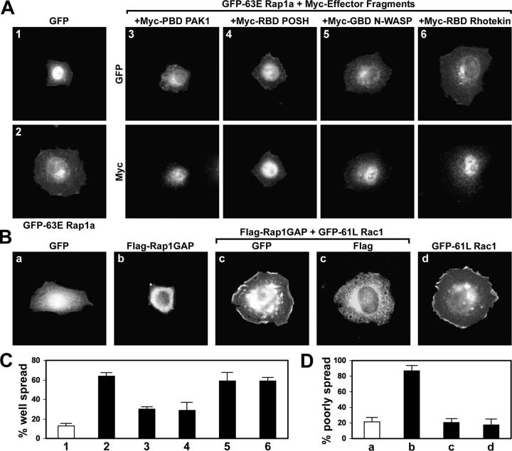
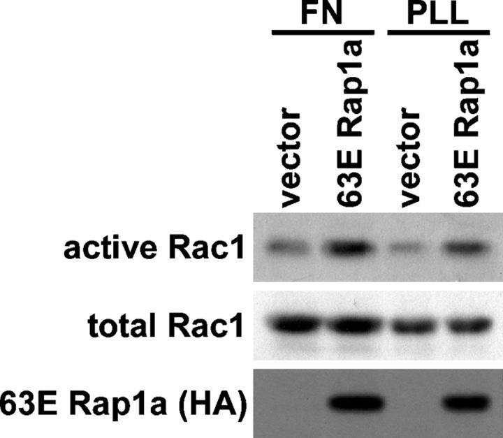
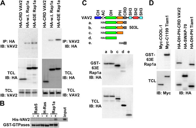
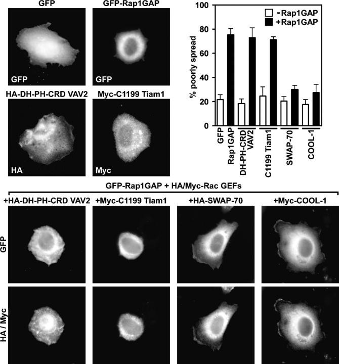
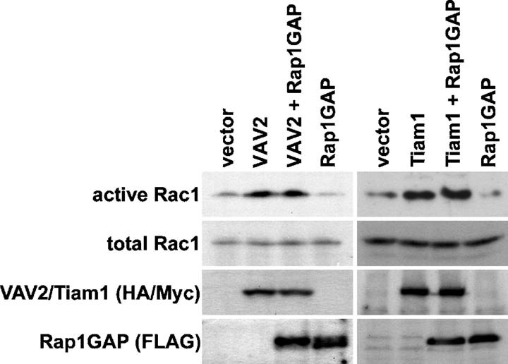
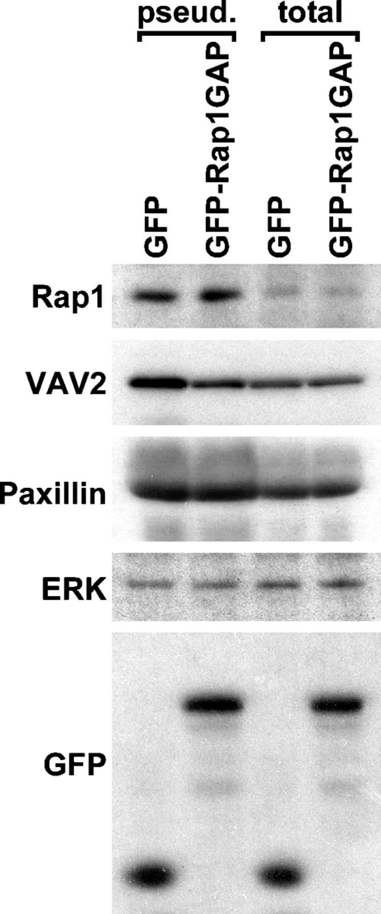
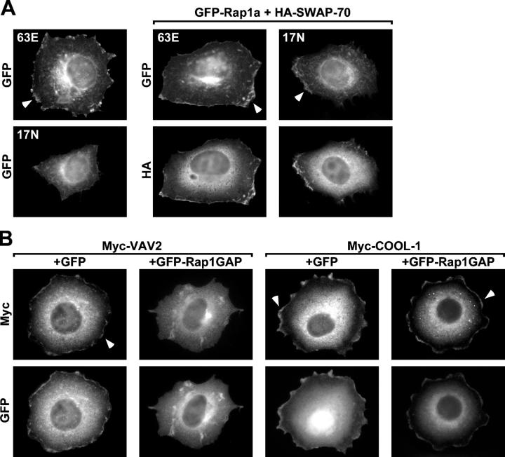
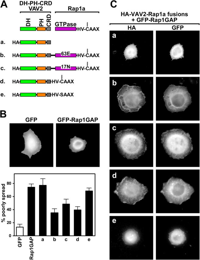
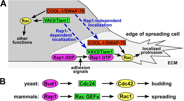
The Ras-related GTPase Rap1 stimulates integrin-mediated adhesion and spreading in various mammalian cell types. Here, we demonstrate that Rap1 regulates cell spreading by localizing guanine nucleotide exchange factors (GEFs) that act via the Rho family GTPase Rac1. Rap1a activates Rac1 and requires Rac1 to enhance spreading, whereas Rac1 induces spreading independently of Rap1. Active Rap1a binds to a subset of Rac GEFs, including VAV2 and Tiam1 but not others such as SWAP-70 or COOL-1. Overexpressed VAV2 and Tiam1 specifically require Rap1 to promote spreading, even though Rac1 is activated independently of Rap1. Rap1 is necessary for the accumulation of VAV2 in membrane protrusions at the cell periphery. In addition, if VAV2 is artificially localized to the cell edge with the subcellular targeting domain of Rap1a, it increases cell spreading independently of Rap1. These results lead us to propose that Rap1 promotes cell spreading by localizing a subset of Rac GEFs to sites of active lamellipodia extension.


Figures

Figure 1.
Figure 1.
Rac1 acts downstream of Rap1 in spreading cells. (A) Rac1 is necessary for cell spreading by active Rap1. HeLa cells were transfected with vectors encoding GFP or constitutively active GFP-63E Rap1a alone or together with inhibitory effector fragments that block signaling downstream from Rho family proteins. Myc-RBD POSH, Myc-GBD N-WASP, and Myc-RBD Rhotekin specifically inhibit signaling from Rac1, Cdc42, and RhoA, respectively. Rac1, Cdc42, and likely other Rho proteins are inhibited by Myc-PBD PAK1. Transfected cells were suspended, plated on fibronectin for 30 min, fixed, and labeled with Myc antibodies. (B) Decreased spreading by Rap1 inactivation is rescued by active Rac1. HeLa cells were transfected with vectors encoding GFP or Flag-Rap1GAP alone or together with constitutively active GFP-61L Rac1, and then allowed to spread on fibronectin for 1 h. (C) Histogram showing the percentage (average plus the SD) of flat, well-spread cells in the experiment described in A. 1, GFP (white bar); 2, GFP-63E Rap1a; 3, GFP-63E Rap1a + Myc-PBD PAK1; 4, GFP-63E Rap1a + Myc-RBD POSH; 5, GFP-63E Rap1a + Myc-GBD N-WASP; 6, GFP-63E Rap1a + Myc-RBD Rhotekin. (D) Histogram showing the percentage (average plus the SD) of refractile, poorly spread cells in the experiment described in B. a, GFP (white bar); b, Flag-Rap1GAP; c, Flag-Rap1GAP + GFP-61L Rac1; d, GFP-61L Rac1.
Figure 2.
Figure 2.
Rap1 activates Rac1. HeLa cells were transfected with an empty vector or a vector encoding constitutively active HA-63E Rap1a. Cells were suspended, plated on fibronectin (FN) or poly-l-lysine (PLL) for 3 h, and lysed, and then active GTP-bound Rac1 was precipitated with GST-PBD PAK1. Precipitates (active) and total cell lysates (total) were then immunoblotted with Rac1 or HA antibodies.
Figure 3.
Figure 3.
Rap1 binds to a subset of Rac GEFs. (A) Rap1 and VAV2 coimmunoprecipitate. HA (left) and VAV2 (right) antibodies were used for immunoprecipitation from serum-starved HeLa cells electroporated with vectors encoding HA-CRD VAV2 (negative control), HA-wild-type Rap1a (mostly GDP bound), or HA-63E Rapla (GTP bound). Immunoprecipitates (IP) and total cell lysates (TCL) were then immunoblotted with HA or VAV2 antibodies. (B) Rap1 binds to VAV2 directly. GTP-loaded GST-Rab5, GST-H-Ras, and GST-Rap1a on glutathione beads were incubated in the absence (−) or presence (+) of bacterially expressed His-DH-PH-CRD VAV2 (input). Beads were washed, and the precipitates were immunoblotted with an anti-His antibody (His-VAV2) or stained with Coomassie blue (GST-GTPases). (C) Rap1 interacts with the DH-PH module of VAV2. GST-63E Rapla was used to pulldown HA-DH-PH-CRD VAV2 (truncation a), HA-DH-PH-CRD W503L VAV2 (b), HA-DH-PH VAV2 (c), HA-DH VAV2 (d), and HA-CRD VAV2 (e) from transiently transfected HeLa cells. Pulldowns (GST-63E Rapla) and total cell lysates were immunoblotted with HA antibodies. (D) Rap1 interacts with a subset of Rac GEFs. GST-63E Rapla was used to pulldown Myc-COOL-1, Myc-C1199 Tiam1, HA-DH-PH-CRD VAV2, HA-SWAP-70, and HA-DH-PH Tiam1 from transiently transfected HeLa cells. Pulldowns and total cell lysates were immunoblotted with Myc (left) or HA (right) antibodies.
Figure 4.
Figure 4.
Rap1 activity is necessary for cell spreading promoted by VAV2 and Tiam1, but not SWAP-70 or COOL-1. HeLa cells were transiently transfected with vectors encoding GFP, GFP-Rap1GAP, HA-DH-PH-CRD VAV2, or Myc-C1199 Tiam1, or cotransfected with vectors encoding GFP-Rap1GAP and HA-DH-PH-CRD VAV2, Myc-C1199 Tiam1, HA-SWAP-70, or Myc-COOL-1. Transfected cells were suspended, plated on fibronectin for 1 h, fixed, and labeled with the HA or Myc antibodies. The histogram shows the percentage (average plus the SD) of refractile, poorly spread cells for each condition in the absence (open bars) or presence (closed bars) of GFP-Rap1GAP.
Figure 5.
Figure 5.
Rap1 activity is dispensable for Rac1 activation by VAV2 and Tiam1. (left) HeLa cells were transfected with an empty vector (vector) or a vector encoding constitutively active HA-DH-PH-CRD VAV2 (VAV2), FLAG-Rap1GAP (Rap1GAP), or both. (right) In separate experiments, cells were transfected with an empty vector or a vector encoding constitutively active Myc-C1199 Tiam1 (Tiam1), FLAG-Rap1GAP, or both. The cells were lysed, and active GTP-bound Rac was precipitated with GST-PBD PAK1. Precipitates (active) and total cell lysates (total Rac1, HA/Myc, FLAG) were immunoblotted with Rac1, HA, Myc, or FLAG antibodies.
Figure 6.
Figure 6.
Rap1 targets VAV2 to matrix-associated membrane protrusions. Pseudopodia and total cell extracts were prepared with Transwell filters from HeLa cells electrotransfected with vectors encoding GFP or GFP-Rap1GAP. Extracts were adjusted for equal content of ERK, a protein found at equal levels in pseudopodia and total cell extracts (Cho and Klemke, 2002). Pseudopodial (pseud.) and total cell (total) extracts were immunoblotted with antibodies for Rap1, VAV2, Paxillin, ERK, or GFP.
Figure 7.
Figure 7.
Rap1 targets VAV2 to circumferential membrane protrusions. (A) Both active and inactive Rap1a localize to membrane protrusions. HeLa cells were transiently transfected with vectors encoding GFP-63E Rapla (63E) or GFP-17N Rap1a (17N) alone (left) or cotransfected with a HA-SWAP-70–encoding vector (right four panels). Transfected cells were suspended, plated on fibronectin for 1 h, fixed, and labeled with HA antibodies. Note that cells expressing 17N Rap1a do not spread, but when spreading is induced with SWAP-70, then 17N Rap1a is detected at the cell periphery. Arrowheads indicate localization of the GFP-Rap1a variants at the cell edge. (B) VAV2, but not COOL-1, requires Rap1 activity to localize to membrane protrusions. HeLa cells were transiently cotransfected with vectors encoding Myc-VAV2 or Myc-COOL-1 and GFP or GFP-Rap1GAP together with HA-SWAP-70. HA-SWAP-70 was cotransfected with the Rac GEFs and GFP vectors to allow Rap1-independent spreading. Transfected cells were treated as in A, labeled with Myc antibodies, and only well-spread cells were analyzed. Arrowheads indicate localization of the GEFs at the cell edge.
Figure 8.
Figure 8.
Rap1-targeted VAV2 fusion proteins function independently of Rap1. (A) Schematic representation of HA epitope-tagged DH-PH-CRD VAV2 (VAV2) fused to full-length 63E Rap1a or 17N Rap1a, consisting of the GTPase domain (GTPase), hypervariable domain (HV), and CAAX box. Also shown are VAV2 fused to the COOH terminus of Rap1a encoding the HV and the prenylated (jagged line) CAAX box. The SAAX mutation abolished membrane targeting by preventing prenylation. HeLa cells were transfected with expression vectors for GFP or GFP-Rap1GAP alone (B) or GFP-Rap1GAP together with HA-VAV2 (a), HA-VAV2-63E Rapla (b), HA-VAV2-17N Rapla (c), HA-VAV2-HV-CAAX (d), or HA-VAV2-HV-SAAX (e) vectors (C). Transfected cells were suspended, plated on fibronectin for 1 h, fixed, and labeled with HA epitope tag antibodies. The histogram shows the percentage (average plus the SD) of refractile, poorly spread cells for each condition.
Figure 9.
Figure 9.
Model: Rap1 regulates morphology by targeting a subset of Rac GEFs to the edge of spreading cells. (A) Only when Rac GEFs are properly targeted do they locally activate Rac, resulting in cell spreading by the formation of productive membrane protrusions adjacent to the ECM. COOL-1 and SWAP-70, two Rac GEFs that do not interact with Rap, are targeted to the protrusive structures by Rap1-independent mechanisms. In contrast, the Rac GEFs VAV2 and Tiam1 are targeted to ECM-associated plasma membranes by binding to active Rap1 (Rap1.GTP) but not inactive Rap1 (Rap1.GDP), following Rap1 activation by adhesion signals. (B) Our proposed model of Rap1 regulation of spreading through Rac GEFs is comparable to the mechanism by which the yeast protein Bud1/Rsr1 controls budding via Cdc24, a Cdc42 GEF.

References

    1. Ardouin, L., M. Bracke, A. Mathiot, S.N. Pagakis, T. Norton, N. Hogg, and V.L. Tybulewicz. 2003. Vav1 transduces TCR signals required for LFA-1 function and cell polarization at the immunological synapse. Eur. J. Immunol. 33:790–797. - PubMed
    1. Asha, H., N.D. de Ruiter, M.G. Wang, and I.K. Hariharan. 1999. The Rap1 GTPase functions as a regulator of morphogenesis in vivo. EMBO J. 18:605–615. - PMC - PubMed
    1. Bishop, A.L., and A. Hall. 2000. Rho GTPases and their effector proteins. Biochem. J. 348:241–255. - PMC - PubMed
    1. Bivona, T.G., H.H. Wiener, I.M. Ahearn, J. Silletti, V.K. Chiu, and M.R. Philips. 2004. Rap1 up-regulation and activation on plasma membrane regulates T cell adhesion. J. Cell Biol. 164:461–470. - PMC - PubMed
    1. Boettner, B., E.E. Govek, J. Cross, and L. Van Aelst. 2000. The junctional multidomain protein AF-6 is a binding partner of the Rap1A GTPase and associates with the actin cytoskeletal regulator profilin. Proc. Natl. Acad. Sci. USA. 97:9064–9069. - PMC - PubMed

Publication types

MeSH terms