. 2004 Nov;78(21):11506-18.
doi: 10.1128/JVI.78.21.11506-11518.2004.

Direct inoculation of simian immunodeficiency virus from sooty mangabeys in black mangabeys (Lophocebus aterrimus): first evidence of AIDS in a heterologous African species and different pathologic outcomes of experimental infection

Affiliations

Direct inoculation of simian immunodeficiency virus from sooty mangabeys in black mangabeys (Lophocebus aterrimus): first evidence of AIDS in a heterologous African species and different pathologic outcomes of experimental infection

Cristian Apetrei et al. J Virol. 2004 Nov.

Abstract

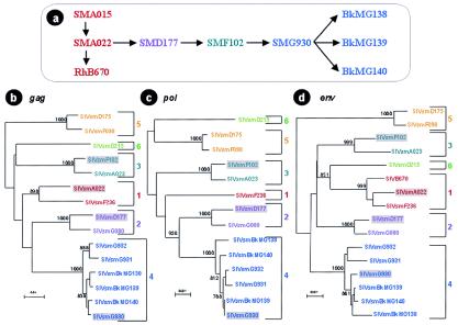
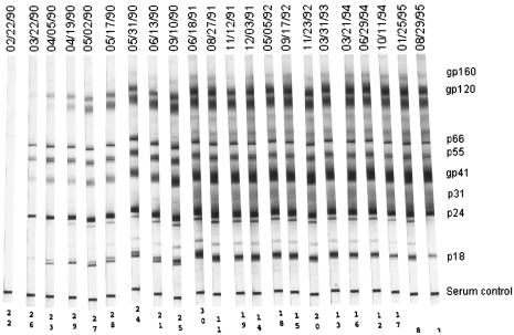
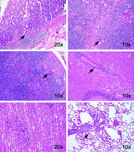
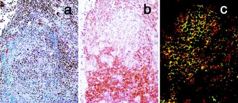
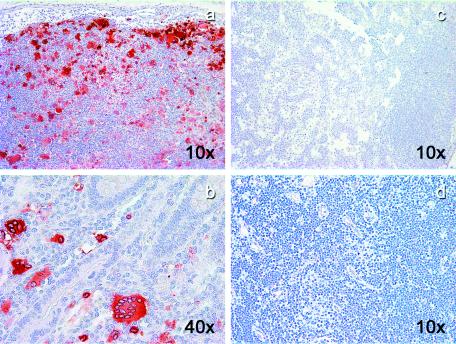
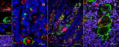
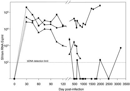
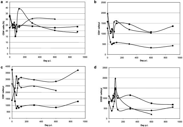
A unique opportunity for the study of the role of serial passage and cross-species transmission was offered by a series of experiments carried out at the Tulane National Primate Research Center in 1990. To develop an animal model for leprosy, three black mangabeys (BkMs) (Lophocebus aterrimus) were inoculated with lepromatous tissue that had been serially passaged in four sooty mangabeys (SMs) (Cercocebus atys). All three BkMs became infected with simian immunodeficiency virus from SMs (SIVsm) by day 30 postinoculation (p.i.) with lepromatous tissue. One (BkMG140) died 2 years p.i. from causes unrelated to SIV, one (BkMG139) survived for 10 years, whereas the third (BkMG138) was euthanized with AIDS after 5 years. Histopathology revealed a high number of giant cells in tissues from BkMG138, but no SIV-related lesions were found in the remaining two BkMs. Four-color immunofluorescence revealed high levels of SIVsm associated with both giant cells and T lymphocytes in BkMG138 and no detectable SIV in the remaining two. Serum viral load (VL) showed a significant increase (>1 log) during the late stage of the disease in BkMG138, as opposed to a continuous decline in VL in the remaining two BkMs. With the progression to AIDS, neopterin levels increased in BkMG138. This study took on new significance when phylogenetic analysis unexpectedly showed that all four serially inoculated SMs were infected with different SIVsm lineages prior to the beginning of the experiment. Furthermore, the strain infecting the BkMs originated from the last SM in the series. Therefore, the virus infecting BkMs has not been serially passaged. In conclusion, we present the first compelling evidence that direct cross-species transmission of SIV may induce AIDS in heterologous African nonhuman primate (NHP) species. The results showed that cross-species-transmitted SIVsm was well controlled in two of three BkMs for 2 and 10 years, respectively. Finally, this case of AIDS in an African monkey suggests that the dogma of SIV nonpathogenicity in African NHP hosts should be reconsidered.


Figures

FIG. 1.
FIG. 1.
(a) Diagram of the serial passage of leprosy lesions between SMs and BkMs. (b to d) Phylogenetic relationships between SIVsm strains infecting the SMs involved in serial passage of M. leprae and of BkMs inadvertently infected with SIVsm during the leprosy experiments. The analysis of gag (640-bp) (b), pol (588-bp) (c), and env (396-bp) (d) gene fragments revealed that all these viruses cluster in different SIVsm lineages and that BkMs were infected with the virus strain naturally infecting SMG930, the final SM in the series. Trees were constructed using the neighbor-joining method with 1,000 replicates. The bootstrap values at the nodes represent the numbers of replicates. Only significant values (>750) are shown. Each lineage is shown in the same color: lineage 1, red; lineage 2, violet; lineage 3, green; lineage 4, blue.
FIG. 2.
FIG. 2.
SIVsm-specific antibody in BkMG138 after inoculation of lepromatous tissues.
FIG. 3.
FIG. 3.
Giant cell disease in BkMG138. Hematoxylin-eosin stain revealed a large number of giant cells in the colon (a), duodenum (b), LNs (c), brain (d), spinal cord (e), and lung (f). The magnifications are shown on each panel.
FIG. 4.
FIG. 4.
Pathology of LNs in BkMG138, which developed AIDS. (a) Atrophic lymphoid follicles and fibrosis (modified trichrome stain); collagen is stained in blue. (b) Immunohistochemistry for CD8+ T cells showing that they predominate in parafollicular areas. (c) Confocal microscopy showing indirect proof of paucity of CD4+ T cells. The CD4+-T-cell depletion is indirectly shown by the high proportion of CD3 (red) and CD8 (green) double-positive cells.
FIG. 5.
FIG. 5.
SIV immunohistochemistry in SIVsm-infected BkMs. Giant cells that appeared in BkMG138 were strongly positive for SIV in both LNs (a) and intestine (b). No evidence of SIV infection could be detected in LNs from BkMG139 (c) and BkMG140 (d).
FIG. 6.
FIG. 6.
Confocal microscopy for triple- and quadruple-fluorescence immunohistochemistry: green (SIV), red (macrophage), blue (T lymphocytes), and magenta (nuclei) stains are evident in BkMG138 diagnosed with giant cell disease. (a) Triple staining of a LN. Large numbers of macrophages and T lymphocytes are infected. (b) Quadruple staining of the intestine from BkMG138. Large numbers of infected cells, mainly macrophages, are visible in the lamina propria. (c) Detail of a quadruple staining of the LN of BkMG138. A large giant cell heavily infected with SIVsm and harboring the macrophage marker HAM56 is visible.
FIG. 7.
FIG. 7.
Dynamics of serum VL in SIVsm-infected BkMs. Due to the sampling schedule, the peak VL could not be measured, and dashed lines covering the interval of days 0 to 30 p.i. are used to reflect this uncertainty. During the follow-up, the VL in BkMG138 (•) showed a significant increase (>1 log) with the progression to AIDS. In contrast, in BkMG139 (▪) and BkMG140 (▴), a tendency to clear viral infection was observed. VL was measured by a bDNA assay (detection limit, 125 eq/ml).
FIG. 8.
FIG. 8.
Dynamics of peripheral immunophenotypic markers during SIVsm infection in BkMs. (a) Percentages of CD4+ cells; (b) numbers of CD4+ cells per microliter; (c) numbers of CD8+ cells per microliter; (d) numbers of CD20+ cells per microliter.Symbols: •, BkMG138; ▪, BkMG139; ▴, BkMG140.

References

    1. Ansari, A. A., N. Onlamoon, P. Bostik, A. E. Mayne, L. Gargano, and K. Pattanapanyasat. 2003. Lessons learnt from studies of the immune characterization of naturally SIV infected sooty mangabeys. Front. Biosci. 8:1030-1050. - PubMed
    1. Apetrei, C., D. L. Robertson, and P. A. Marx. 2004. The history of SIVs and AIDS: epidemiology, phylogeny and biology of isolates from naturally infected non-human primates (NHP) in Africa. Front. Biosci. 9:225-254. - PubMed
    1. Ayouba, A., P. Mauclere, P. M. Martin, P. Cunin, J. Mfoupouendoun, B. Njinku, S. Souquieres, and F. Simon. 2001. HIV-1 group O infection in Cameroon, 1986 to 1998. Emerg. Infect. Dis. 7:466-467. - PMC - PubMed
    1. Balfe, P., P. Simmonds, C. A. Ludlam, J. O. Bishop, and A. J. Brown. 1990. Concurrent evolution of human immunodeficiency virus type 1 in patients infected from the same source: rate of sequence change and low frequency of inactivating mutations. J. Virol. 64:6221-6233. - PMC - PubMed
    1. Barnet, W. S., M. K. Krishna, B. G. Herndier, and J. A. Levy. 1994. An AIDS-like condition induced in baboons by HIV-2. Science 266:642-646. - PubMed

Publication types

MeSH terms

LinkOut - more resources