. 2004 Nov;78(21):11551-62.
doi: 10.1128/JVI.78.21.11551-11562.2004.

Intracellular localization and protein interactions of the gene 1 protein p28 during mouse hepatitis virus replication

Affiliations

Intracellular localization and protein interactions of the gene 1 protein p28 during mouse hepatitis virus replication

Sarah M Brockway et al. J Virol. 2004 Nov.

Abstract

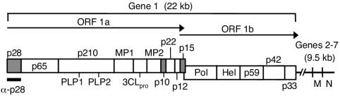
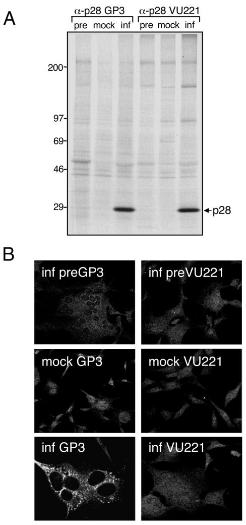
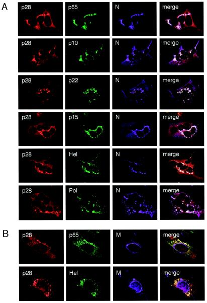
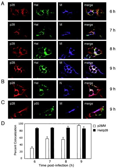
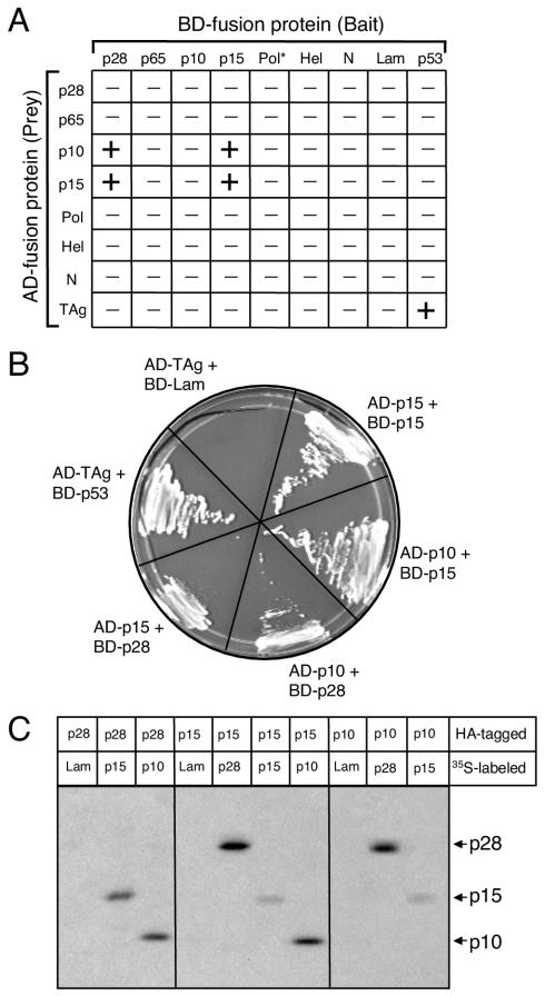
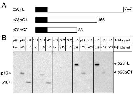
Coronaviruses encode the largest replicase polyprotein of any known positive-strand RNA virus. Replicase protein precursors and mature products are thought to mediate the formation and function of viral replication complexes on the surfaces of intracellular double-membrane vesicles. However, the functions of only a few of these proteins are known. For the coronavirus mouse hepatitis virus (MHV), the first proteolytic processing event of the replicase polyprotein liberates an amino-terminal 28-kDa product (p28). While previous biochemical studies have suggested that p28 is associated with viral replication complexes, the intracellular localization and interactions of p28 with other proteins during the course of MHV replication have not been defined. We used immunofluorescence confocal microscopy to show that p28 localizes to viral replication complexes in the cytoplasm during early times postinfection. However, at late times postinfection, p28 localizes to sites of M accumulation distinct from the replication complex. Furthermore, by yeast two-hybrid and coimmunoprecipitation analyses, we demonstrate that p28 specifically binds to p10 and p15, two coronavirus replicase proteins of unknown function. Deletion mutagenesis experiments determined that the carboxy terminus of p28 is not required for its interactions with p10 and p15. These results suggest that p28 may play a part at the replication complex by interacting with p10 and p15. Moreover, our findings highlight a potential role for p28 at virion assembly sites.


Figures

FIG. 1.
FIG. 1.
MHV genome organization. The diagram shows the organization of the 22-kb MHV-A59 gene 1 (replicase gene) and the locations of genes 6 and 7, encoding the structural proteins M and N, respectively. Gene 1 is composed of two open reading frames (ORF1a and ORF1b). The ORF1a-ORF1b fusion replicase polyprotein is shown, with mature replicase proteins represented as boxes. Proteins with confirmed or predicted functions include two papain-like proteinases (PLP1 and PLP2), the 3C-like proteinase (3CLpro), two transmembrane proteins (MP1 and MP2), the RNA-dependent RNA polymerase (Pol), and the RNA helicase (Hel). All other proteins are labeled based on their molecular masses in kilodaltons. The gray boxes represent replicase proteins of interest in the present study: the amino-terminal cleavage product, p28, and two carboxy-terminal ORF1a proteins, p10 and p15. The region of the gene 1 polyprotein (amino acids M1 to G247) used to generate α-p28 guinea pig (GP3) and rabbit (VU221) polyclonal antisera is indicated.
FIG. 2.
FIG. 2.
Detection of p28 by immunoprecipitation and immunofluorescence. (A) Immunoprecipitation of p28. Lysates from either mock-infected (mock) or MHV-infected (inf) radiolabeled DBT cells were immunoprecipitated using antiserum raised against p28 (GP3 or VU221) in buffer C as described in Materials and Methods. Lysate from infected cells was also immunoprecipitated using preimmune sera from the same animal (pre). Bands corresponding to p28 are indicated on the right of the gel, and molecular mass standards (in kilodaltons) are shown on the left. (B) Immunofluorescence analysis using p28 antisera. DBT cells grown on glass coverslips were either mock infected or infected with MHV for 6 h, fixed and permeabilized with 100% methanol, and incubated with GP3, VU221, or preimmune sera using an indirect immunofluorescence assay. The cells were imaged using a Zeiss LSM 510 confocal microscope at 546 nm. The images are single confocal slices obtained using a 40× objective and are representative of the cell population. Multinucleated cells are a cytopathic effect of MHV replication.
FIG. 3.
FIG. 3.
Triple-label immunofluorescent images of MHV-infected DBT cells at 6 h p.i. (A) p28 colocalizes with MHV replication complexes. DBT cells grown on glass coverslips were infected with MHV for 6 h, fixed and permeabilized with 100% methanol, and incubated with antibodies against p28 (GP3) (red), various MHV replicase proteins (green), and N (purple). Colocalization of all three colors is shown as white pixels. The cells were imaged using a Zeiss LSM 510 confocal microscope at 488, 546, and 633 nm. The images are single confocal slices obtained using a 40× objective and are representative of the cell population. (B) p28 is distinct from sites of M accumulation. Cells were incubated with antibodies against p28 (GP3) (red), replicase protein p65 or Hel (green), and the viral M protein (purple) as a marker of virion assembly sites. Colocalization of red and green is shown as yellow pixels.
FIG. 4.
FIG. 4.
Time course of p28 localization during MHV infection. MHV-infected DBT cells grown on glass coverslips were fixed at the times p.i. shown on the right of the images and stained with antibodies against viral replicase and structural proteins. (A) p28 colocalizes with Hel over the course of MHV infection. Individual coverslips were stained with antibodies against p28 (GP3) (red), Hel (green), and M (purple), a marker for sites of virion assembly. (B) p28 is associated with Hel and N at 9 h p.i. Coverslips from the 9-h time point were stained with antibodies against p28 (GP3) (red), Hel (green), and N (purple). Colocalization of all three colors is shown as white pixels. (C) p28 is distinct from replication complexes at 9 h p.i. Coverslips from the 9-h time point were incubated with antibodies against p28 (GP3) (red), M (purple), and the replicase protein p65 (green) as a marker for replication complex staining at late times p.i. Colocalization of red and purple is shown as pink pixels. (D) Quantitation of percent colocalization of p28, Hel, and M. Percent colocalization was determined using Metamorph Imaging software. The background fluorescence was determined empirically by staining mock-infected cells with immune sera. The lower-limit threshold was set to exclude pixels below background. The upper-limit level was set to exclude saturated pixels. For colocalization measurements of each protein pair at each time point, three independent images (∼5 cells per image) were acquired and processed. The error bars indicate standard deviations.
FIG. 5.
FIG. 5.
Interactions between MHV proteins using yeast two-hybrid and coimmunoprecipitation assays. (A) Pairwise cotransformations. Various MHV replicase proteins and N were expressed as either bait (fused to the GAL4 BD) or prey (fused to the GAL4 AD) in pairwise combinations using the yeast two-hybrid system. Yeast expressing both bait and prey fusion proteins was scored for the capacity to grow on medium lacking Trp, Leu, Ade, and His and by blue-white screening in the presence of X-α-Gal. In the matrix shown, − indicates lack of growth and + indicates both growth and blue color. The interaction between TAg and p53 served as a positive control for the experiment; TAg and lamin C (Lam) served as a negative control. The asterisk indicates that BD-Pol activates the reporter gene MEL1. (B) Interactions among p28, p10, and p15. Single yeast colonies containing the indicated bait and prey plasmids were streaked onto medium lacking Trp, Leu, Ade, or His to confirm growth. (C) Coimmunoprecipitation of in vitro-expressed proteins. Proteins were translated, using reticulocyte lysate, as fusions to c-Myc epitope tags in the presence of [35S]methionine or as fusions to HA epitope tags in the absence of radiolabel. Equal amounts of radiolabeled and nonradiolabeled lysates were combined, and the proteins were immunoprecipitated using rabbit polyclonal α-HA antiserum. Interacting 35S-labeled proteins were resolved in an SDS-12% polyacrylamide gel and analyzed by fluorography. The identities of coprecipitating proteins are shown on the right of the gel.
FIG. 6.
FIG. 6.
The carboxy terminus of p28 is not required for interactions with p10 and p15. (A) p28 deletion mutagenesis. A schematic of full-length 247-amino-acid p28 (p28FL) is shown with the two carboxy-terminal truncation mutants, p28ΔC1 and p28ΔC2, below p28FL. The number of p28 amino acid residues remaining is indicated to the right of each protein. The c-Myc or HA epitope tag is represented as a black box fused to the amino terminus of each p28 protein and is not drawn to scale. (B) Coimmunoprecipitation of in vitro-expressed proteins. The proteins were translated, using reticulocyte lysates, as fusions to c-Myc epitope tags in the presence of [35S]methionine or as fusions to HA epitope tags in the absence of radiolabel. Equal amounts of radiolabeled and nonradiolabeled lysates were combined, and the proteins were immunoprecipitated using rabbit polyclonal α-HA antiserum. Interacting 35S-labeled proteins were resolved in an SDS-12% polyacrylamide gel and analyzed by fluorography. The identities of coprecipitating proteins are shown on the right of the gel.
FIG. 7.
FIG. 7.
Coimmunoprecipitation of proteins from MHV-infected cell lysates. Mock-infected or MHV-infected cells were radiolabeled for 3 h at 5 h p.i., and cytoplasmic extracts were immunoprecipitated using buffer A (1% NP-40 buffer), buffer B (0.1% SDS buffer), or buffer C (1% SDS plus preboiling lysate) and antiserum against replicase protein p28, p10, or p15. Buffer A was used for control immunoprecipitations, including those with mock-infected lysate immunoprecipitated with immune sera (M) and infected lysate immunoprecipitated with preimmune sera (pre). Proteins were analyzed following SDS-PAGE in 5 to 18% polyacrylamide gradient gels and by fluorography. Molecular mass markers (in kilodaltons) are shown on the left, and the proteins of interest are indicated on the right of the gels. (A) α-p28 (GP3). (B) α-p10. (C) α-p28 (VU221). (D) α-p15. (E) p28 coprecipitating proteins have the same mobility as p10 and p15. Radiolabeled infected cell lysate was immunoprecipitated with antiserum specific for p28 (GP3 or VU221), p10 (α-p10), or p15 (α-p15) using buffer A conditions. The proteins were analyzed following SDS-PAGE in a 12% polyacrylamide gradient gel and fluorography. The identities of precipitated proteins are indicated to the right of the gels.

References

    1. Baric, R. S., G. W. Nelson, J. O. Fleming, R. J. Deans, J. G. Keck, N. Casteel, and S. A. Stohlman. 1988. Interactions between coronavirus nucleocapsid protein and viral RNAs: implications for viral transcription. J. Virol. 62:4280-4287. - PMC - PubMed
    1. Bi, W., J. D. Pinon, S. Hughes, P. J. Bonilla, K. V. Holmes, S. R. Weiss, and J. L. Leibowitz. 1998. Localization of mouse hepatitis virus open reading frame 1A derived proteins. J. Neurovirol. 4:594-605. - PubMed
    1. Bonilla, P. J., A. E. Gorbalenya, and S. R. Weiss. 1994. Mouse hepatitis virus strain A59 RNA polymerase gene ORF 1a: heterogeneity among MHV strains. Virology 198:736-740. - PMC - PubMed
    1. Bost, A. G., R. H. Carnahan, X. T. Lu, and M. R. Denison. 2000. Four proteins processed from the replicase gene polyprotein of mouse hepatitis virus colocalize in the cell periphery and adjacent to sites of virion assembly. J. Virol. 74:3379-3387. - PMC - PubMed
    1. Bost, A. G., E. Prentice, and M. R. Denison. 2001. Mouse hepatitis virus replicase protein complexes are translocated to sites of M protein accumulation in the ERGIC at late times of infection. Virology 285:21-29. - PMC - PubMed

Publication types

Substances

LinkOut - more resources