. 2004 Nov;78(21):11739-50.
doi: 10.1128/JVI.78.21.11739-11750.2004.

Lv1 inhibition of human immunodeficiency virus type 1 is counteracted by factors that stimulate synthesis or nuclear translocation of viral cDNA

Affiliations

Lv1 inhibition of human immunodeficiency virus type 1 is counteracted by factors that stimulate synthesis or nuclear translocation of viral cDNA

Lionel Berthoux et al. J Virol. 2004 Nov.

Abstract

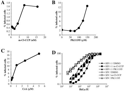
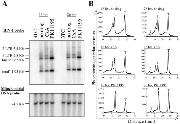
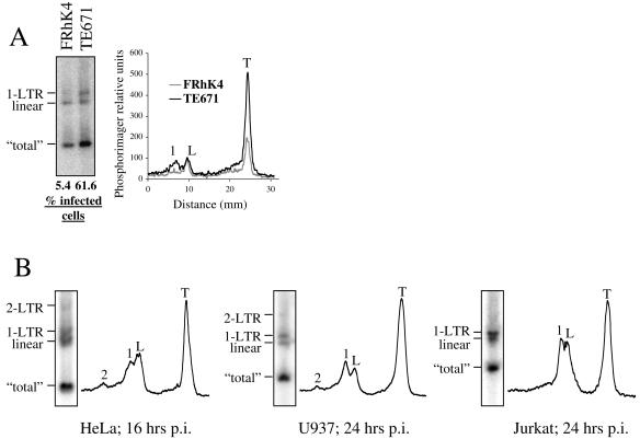
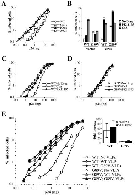
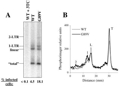
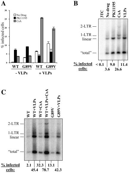
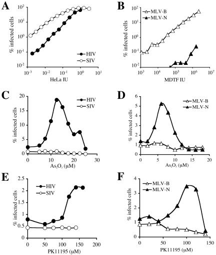
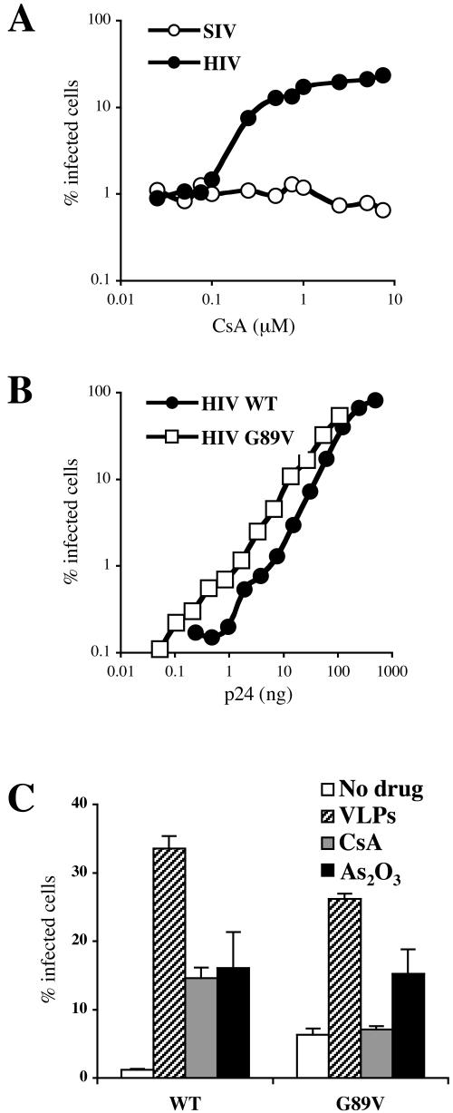
Human immunodeficiency virus type 1 (HIV-1) cDNA synthesis is inhibited in cells from some nonhuman primates by an activity called Lv1. Sensitivity to restriction by Lv1 maps to a region of the HIV-1 CA required for interaction with the cellular protein cyclophilin A. A similar antiviral activity in mammalian cells, Ref1, inhibits reverse transcription of murine leukemia virus (MLV), but only with viral strains bearing N-tropic CA. Disruption of the HIV-1 CA-cyclophilin A interaction inhibits Lv1 restriction in some cells and, paradoxically, seems to render HIV-1 sensitive to Ref1. Lv1 and Ref1 activities are overcome by high-titer infection and are saturable with nonreplicating, virus-like particles encoded by susceptible viruses. Two compounds that disrupt mitochondrial membrane potential, As(2)O(3) and m-Cl-CCP, reduce Ref1 activity. Here we show that these drugs, as well as a third compound with similar effects on mitochondria, PK11195, attenuate Lv1 activity in rhesus macaque and African green monkey cells. Effects of PK11195 and virus-like particles on HIV-1 infectivity in these cells were largely redundant, each associated with increased HIV-1 cDNA. Comparison of acutely infected macaque and human cells suggested that, in addition to effects on cDNA synthesis, Lv1 inhibits the accumulation of nuclear forms of HIV-1 cDNA. Disruption of the HIV-1 CA-cyclophilin A interaction caused a minimal increase in total viral cDNA but increased the proportion of viral cDNA in the nucleus. Consistent with a model in which Lv1 inhibits both synthesis and nuclear translocation of HIV-1 cDNA, complete suppression of macaque or African green monkey Lv1 was achieved by the additive effect of factors that stimulate both processes.


Figures

FIG. 1.
FIG. 1.
(A to C) m-Cl-CCP, PK11195, and CsA each counteract Lv1 activity in rhesus macaque cells. FRhK4 cells were infected with VSV-G-pseudotyped HIV-1NL-GFP in the presence of the indicated concentrations of m-Cl-CCP (A), PK11195 (B), or CsA (C). Supernatant was replaced with drug-free medium 16 h postinfection, and the percentage of cells expressing GFP was determined 2 days later by flow cytometry. (D) FRhK4 cells were infected with VSV-G-pseudotyped virus, either HIV-1NL-GFP or SIVMAC239-GFP, after normalizing the titer of the two viruses on HeLa cells (HeLa IU). Where indicated, infections were performed in the presence of 100 μM PK11195 or 10 nM m-Cl-CCP. GFP expression was analyzed by flow cytometry at 48 h.
FIG. 2.
FIG. 2.
Effect of CsA and PK11195 on HIV-1 cDNA synthesis. (A) FRhK4 cells were infected with VSV-G-pseudotyped HIV-1 for 16 or 26 h in the presence of PK11195, CsA, or the reverse transcriptase inhibitor 3TC. Total cellular DNA was extracted, digested with XhoI, MscI, and DpnI, and processed for Southern blotting. The membrane was probed with an HIV-1-specific DNA probe (top) and then stripped and reprobed for human mitochondrial DNA (bottom). Viral DNA species are identified on the left of the blot. The total band corresponds to a DNA fragment that is part of all viral DNA species, including proviral DNA. (B) Plots of signal intensity for the indicated lanes from panel A, as determined with a phosphorimager. The main viral DNA species are indicated above the peaks. 2, 2-LTR circle; 1, 1-LTR circle; L, linear; T, total. Note differences in scales between panels.
FIG. 3.
FIG. 3.
Southern analysis of HIV-1 cDNA synthesis in rhesus macaque and human cell lines. (A) Comparison of HIV-1 cDNA species in FRhK4 and human TE671 cells infected for 16 h with the same quantity of VSV-G-pseudotyped HIV-1NL-GFP. An aliquot of each sample was maintained in culture for 2 days, and the percentage of GFP-positive cells was determined by flow cytometry. (B) Southern analysis of HIV-1 cDNA synthesis in HeLa, U937, and Jurkat T cells, as described in the legend for Fig. 2.
FIG. 4.
FIG. 4.
Disruption of the interaction between CypA and CA increases HIV-1 infectivity in rhesus macaque cells. FRhK4 cells were infected with GFP-transducing HIV-1 vectors (A and B) or viruses (B to E), either wild type or bearing the indicated CA mutations, in the presence of PK11195 or CsA (B to D) or in the presence of VLPs (E). Supernatant was replaced with drug-free medium 16 h later, and the percentage of GFP-expressing cells was determined 2 days later. The inset in panel E shows the average magnitude increase in infectivity due to addition of wild-type or G89V VLPs, when the percentage of infected cells in the absence of VLPs was less than 2%.
FIG. 5.
FIG. 5.
Effect of the G89V CA mutant on HIV-1 cDNA synthesis. FRhK4 cells were infected for 16 h with wild-type or G89V mutant HIV-1NL-GFP. The two viruses were normalized by p24 ELISA. (A) Cells were trypsinized, and total DNA was extracted and processed for Southern blotting as described in the legend for Fig. 2. Similar amounts of DNA were loaded in each lane, as demonstrated with a probe against mitochondrial DNA (data not shown). As a control, cells were infected with wild-type virus in the presence of 3TC. The main viral DNA species are identified on the left of the blot. One-twentieth of each culture was set aside to determine the percentage of cells expressing GFP, as indicated at the bottom of each lane. (B) Signals in each lane of panel A were quantitated by a phosphorimager as described in the legend for Fig. 2.
FIG. 6.
FIG. 6.
HIV-1 cDNA synthesis after infection of macaque cells is increased in the presence of VLPs. (A) Wild-type or G89V mutant HIV-1NL-GFP (normalized by p24 ELISA) were used to infect FRhK4 cells in the presence of wild-type HIV-1 VLPs, PK11195, or CsA as indicated. The percentage of cells expressing GFP was determined 2 days later by flow cytometry. (B) FRhK4 cells were infected for 16 h with VSV-G-pseudotyped HIV-1 in the presence of PK11195, CsA, VLPs, or 3TC, as indicated. Cells were processed for Southern blotting as described for Fig. 2. (C) FRhK4 cells were infected with wild-type or G89V HIV-1NL-GFP and treated with either VLPs or CsA (5 μM) or a combination of the two. HIV-1 cDNA was detected by Southern blotting as described above. The percentage of GFP-expressing cells was determined as above and is indicated at the bottom of the figure.
FIG. 7.
FIG. 7.
As2O3 and PK11195 counteract Ref1 and Lv1 activity in African green monkey cells. (A) HIV-1NL-GFP and SIVMAC239-GFP were used to infect Vero cells with multiple doses of virus. The two virus stocks were normalized by determining infectious units on HeLa cells. (B) B-tropic and N-tropic GFP-expressing MLV vectors were used to infect Vero cells. Virus doses are expressed as infectious units on nonrestrictive MDTF cells. Vero cells were infected with HIV-1NL-GFP and SIVMAC239-GFP (C and E) or MLV-N and MLV-B (D and F) in the presence of the indicated concentrations of As2O3 (C and D) or PK11195 (E and F). In all cases, the percentage of cells expressing GFP at 48 h postinfection was determined by flow cytometry.
FIG. 8.
FIG. 8.
Effect of CsA or the HIV-1 CA G89V mutation on Lv1 activity in African green monkey cells. (A) HIV-1NL-GFP and SIVMAC239-GFP were used to infect Vero cells in the presence of the indicated concentrations of CsA. (B and C) Wild-type and G89V mutant HIV-1NL-GFP (normalized by ELISA for p24) were used to infect Vero cells at multiple virus doses (B) or at a single dose in the presence of HIV-1 VLPs, PK11195, or CsA (C). In each case, the percentage of cells expressing GFP was determined 2 days postinfection by flow cytometry.

References

    1. Aberham, C., S. Weber, and W. Phares. 1996. Spontaneous mutations in the human immunodeficiency virus type 1 gag gene that affect viral replication in the presence of cyclosporins. J. Virol. 70:3536-3544. - PMC - PubMed
    1. Adachi, A., H. E. Gendelman, S. Koenig, T. Folks, R. Willey, A. Rabson, and M. A. Martin. 1986. Production of acquired immunodeficiency syndrome-associated retrovirus in human and nonhuman cells transfected with an infectious molecular clone. J. Virol. 59:284-291. - PMC - PubMed
    1. Armstrong, J. S., K. K. Steinauer, J. French, P. L. Killoran, J. Walleczek, J. Kochanski, and S. J. Knox. 2001. Bcl-2 inhibits apoptosis induced by mitochondrial uncoupling but does not prevent mitochondrial transmembrane depolarization. Exp. Cell Res. 262:170-179. - PubMed
    1. Bell, P., L. J. Montaner, and G. G. Maul. 2001. Accumulation and intranuclear distribution of unintegrated human immunodeficiency virus type 1 DNA. J. Virol. 75:7683-7691. - PMC - PubMed
    1. Berthoux, L., C. Pechoux, and J. L. Darlix. 1999. Multiple effects of an anti-human immunodeficiency virus nucleocapsid inhibitor on virus morphology and replication. J. Virol. 73:10000-10009. - PMC - PubMed

Publication types