

Transmission study of Andes hantavirus infection in wild sigmodontine rodents
- PMID: 15479837
- PMCID: PMC523238
- DOI: 10.1128/JVI.78.21.11972-11979.2004
Transmission study of Andes hantavirus infection in wild sigmodontine rodents
Abstract
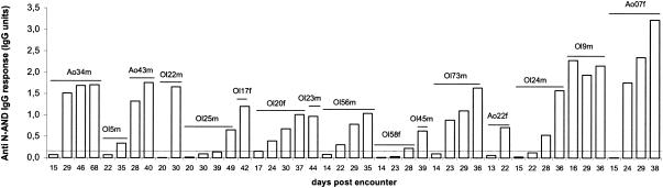
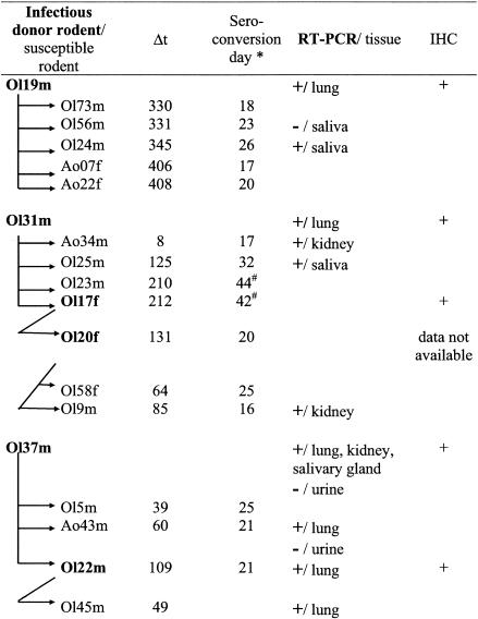
Our study was designed to contribute to an understanding of the timing and conditions under which transmission of Andes hantavirus in Oligoryzomys longicaudatus reservoir populations takes place. Mice were caged in test habitats consisting of steel drums containing holding cages, where seronegative rodents were exposed to wild seropositive individuals by freely sharing the same cage or being separated by a wire mesh. Tests were also performed for potential viral transmission to mice from excrement-tainted bedding in the cages. Andes virus transmitted efficiently; from 130 attempts with direct contact, 12.3% resulted in virus transmission. However, if we consider only those rodents that proved to be infectious, from 93 attempts we obtained 16 infected animals (17.2%). Twelve of them resulted from intraspecies O. longicaudatus encounters where male mice were differentially affected and 4 resulted from O. longicaudatus to Abrothrix olivaceus. Experiments using Abrothrix longipilis as receptors were not successful. Transmission was not observed between wire mesh-separated animals, and mice were not infected from excrement-tainted bedding. Bites seemed not to be a requisite for oral transmission. Genomic viral RNA was amplified in two out of three saliva samples from seropositive rodents, but it was not detected in urine samples obtained by vesicle puncture from two other infected rodents. Immunohistochemistry, using antibodies against Andes (AND) hantavirus proteins, revealed strong reactions in the lung and salivary glands, supporting the possibility of oral transmission. Our study suggests that AND hantavirus may be principally transmitted via saliva or saliva aerosols rather than via feces and urine.
Figures

References
-
- Armstrong, L. R., S. R. Zaki, M. J. Goldoft, R. L. Todd, A. S. Khan, R. F. Khabbaz, T. G. Ksiazek, and C. J. Peters. 1995. Hantavirus pulmonary syndrome associated with entering or cleaning rarely used, rodent-infested structures. J. Infect. Dis. 172:1166. - PubMed
-
- Cadiz, R. 2000. Estudio de la seroprevalencia de hantavirus en reservorios silvestres en distintos habitat de la décima región y análisis de su comportamiento temporal en poblaciones de roedores del fundo experimental San Martín. Tesis de grado, medico veterinario. Universidad Austral de Chile, Valdivia.
-
- Cantoni, G., P. Padula, G. Calderon, J. Mills, E. Herrero, P. Sandoval, V. Martinez, N. Pini, and E. Larrieu. 2001. Seasonal variation in prevalence of antibody to hantaviruses in rodents from southern Argentina. Trop. Med. Int. Health 6:811-816. - PubMed
-
- Childs, J. E., T. G. Ksiazek, C. F. Spiropoulou, J. W. Krebs, S. Morzunov, G. O. Maupin, K. L. Gage, P. E. Rollin, J. Sarisky, R. E. Enscore, et al. 1994. Serologic and genetic identification of Peromyscus maniculatus as the primary rodent reservoir for a new hantavirus in the southwestern United States. J. Infect. Dis. 169:1271-1280. - PubMed
Publication types
MeSH terms
Substances
LinkOut - more resources
Full Text Sources
Other Literature Sources
Medical
Molecular Biology Databases
