. 2004 Nov;78(21):11980-7.
doi: 10.1128/JVI.78.21.11980-11987.2004.

Infection of specific dendritic cells by CCR5-tropic human immunodeficiency virus type 1 promotes cell-mediated transmission of virus resistant to broadly neutralizing antibodies

Affiliations

Infection of specific dendritic cells by CCR5-tropic human immunodeficiency virus type 1 promotes cell-mediated transmission of virus resistant to broadly neutralizing antibodies

Lakshmanan Ganesh et al. J Virol. 2004 Nov.

Abstract

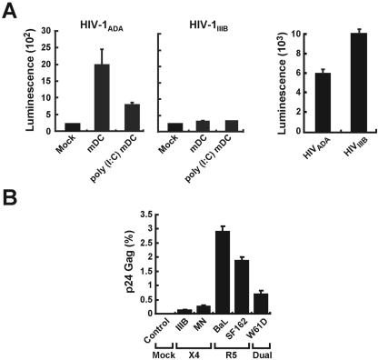
The tropism of human immunodeficiency virus type 1 for chemokine receptors plays an important role in the transmission of AIDS. Although CXCR4-tropic virus is more cytopathic for T cells, CCR5-tropic strains are transmitted more frequently in humans for reasons that are not understood. Phenotypically immature myeloid dendritic cells (mDCs) are preferentially infected by CCR5-tropic virus, in contrast to mature mDCs, which are not susceptible to infection but instead internalize virus into a protected intracellular compartment and enhance the infection of T cells. Here, we define a mechanism to explain preferential transmission of CCR5-tropic viruses based on their interaction with mDCs and sensitivity to neutralizing antibodies. Infected immature mDCs differentiated normally and were found to enhance CCR5-tropic but not CXCR4-tropic virus infection of T cells even in the continuous presence of neutralizing antibodies. Infectious synapses also formed normally in the presence of such antibodies. Infection of immature mDCs by CCR5-tropic virus can therefore establish a pool of infected cells that can efficiently transfer virus at the same time that they protect virus from antibody neutralization. This property of DCs may enhance infection, contribute to immune evasion, and could provide a selective advantage for CCR5-tropic virus transmission.


Figures

FIG. 1.
FIG. 1.
HIV infection of mDCs. (A) Direct viral infection by replication-defective HIV reporter viruses of mDCs (left and middle) and T cells (A3R5; right). Untreated mDCs and poly(I-C)-treated mDCs (3 × 10⁴ cells) were infected with either HIV-1ADA (left) or HIV-1IIIB (middle) (25 ng of p24) for 2 h. A3R5 T cells (3 × 10⁴ cells) were also infected with HIV-1ADA or HIV-1IIIB (25 ng of p24) for 2 h (right). Cells washed three times and maintained in cell culture were collected 2 days later for luciferase assay. (B) Direct viral infection of mDCs by CXCR4-tropic (X4), CCR5-tropic (R5), and dualtropic (Dual), HIV-1 isolates. mDCs (3 × 10⁵/well) isolated from a single donor elutriated monocytes (see Materials and Methods) were mock infected or infected with the indicated HIV-1 strains for 12 h, washed, incubated for 48 h, and analyzed by fluorescence-activated cell sorting for intracellular p24 after gating to exclude EMA and to select for CD11c cells.
FIG. 2.
FIG. 2.
Infection with HIV-1BaL does not prevent maturation of mDCs by poly(I-C). mDCs (3 × 10⁵/well) isolated from a single donor elutriated monocytes (see Materials and Methods) were infected with HIV-1BaL (2 μg of p24/ml). The control groups were treated with AZT (10 μM; left top and bottom panels) to prevent HIV replication. One set of control and HIV-infected cells (left lower panel, second lower panel) were matured 48 h after infection with poly(I-C) (50 μg/ml). To determine the relationship between infection and maturation, CD80 and CD40 expression (in separate experiments) were assessed in the Gag+ population compared to the Gag population in untreated (right upper) and poly(I-C)-treated (right lower) groups of DCs.
FIG. 3.
FIG. 3.
Effect of antibody exposure on HIV-1 trans-infection. (A) HIV-1BaL-infected immature DCs transfer the virus more effectively to T cells upon maturation. mDCs (3 × 10⁵/well) isolated from a single donor (elutriated monocytes) were mock infected or infected with HIV-1BaL (2 μg of p24/ml). One set of mock-infected and HIV-infected mDCs was matured 48 h after infection with poly(I-C) (50 μg/ml). After 48 h, primary PHA-IL-2-stimulated autologous CD4+ T cells were added to both mock-infected and HIV-1-infected immature and mature mDCs, followed by incubation in the presence of indinavir (1 μM) for another 48 h. Cells were then assayed for intracellular Gag by flow cytometry. (B) Poly(I-C)-treated mDCs were infected with HIV-1BaL for 2 h, washed five times, and incubated with the indicated concentrations of 2F5 (left) or b12 (right) and primary PHA-IL-2-stimulated autologous CD4+ T cells for 30 min or 48 h. In the control group, the virus was preincubated with antibody for 30 min prior to infection of CD4+ T cells for 2 h. Cells were maintained in indinavir (1 μM), and p24 Gag in CD3+ CD11c cells was assayed by fluorescence-activated cell sorting 48 h later. Percent neutralization was defined as the reduction in the number of p24-Ag-positive cells compared to the number in control wells with no antibody (29).
FIG. 4.
FIG. 4.
mDCs infected with HIV-1BaL confer resistance to antibody neutralization. Wild-type HIV-1IIIB, HIV-1BaL, or HIV-189.6 (12 ng of p24) was exposed to anti-gp120 antibody, b12 (50 μg/ml), for 60 min before infection of T cells (A) or mDCs (B) (4 × 10⁴ cells each) for 2 h. (C) Alternatively, these viruses were used to infect mDCs (4 × 10⁴ cells each) for 2 h. Cells were washed five times to remove virus and either incubated with T cells alone (105 cells) (B) or treated with b12 (50 μg/ml) and T cells (105 cells) (C). b12 was added every 60 h and left in the DC-T-cell mixture for the duration of the experiments (C). At the appropriate time, cell supernatants were collected, and p24 ELISA was performed as instructed by the manufacturer (Coulter). The data are representative of duplicate experiments.
FIG. 5.
FIG. 5.
Confocal imaging of the infectious synapse and implications of mDC transmission of CCR5-tropic HIV to T cells. (A) Neutralizing b12 antibody does not inhibit formation of infectious synapses. Virus infection of mDCs was performed with a Vpr-GFP-labeled virus vector described previously (31) and assessed by confocal microscopy (see Materials and Methods; control versus b12, P = 0.5) with representative cells. The arrow indicates a concentrated region of labeled virus. (B) Model of HIV infection of immature mDCs, internalization, and transfer by mature mDCs that confers resistance to antibody neutralization.

References

    1. Abrahamsen, T. G., C. S. Carter, E. J. Read, M. Rubin, H. G. Goetzman, E. F. Lizzio, Y. L. Lee, P. A. Pizzo, and T. Hoffman. 1991. Stimulatory effect of counterflow centrifugal elutriation in large-scale separation of peripheral blood monocytes can be reversed by storing the cells at 37°C. J. Clin. Apheresis 6:48-53. - PubMed
    1. Alvarez, C. P., F. Lasala, J. Carrillo, A. L. Corbi, A. L. Corbi, and R. Delgado. 2002. C-type lectins DC-SIGN and L-SIGN mediate cellular entry by Ebola virus in cis and in trans. J. Virol. 76:6841-6844. - PMC - PubMed
    1. Ayehunie, S., E. A. Garcia-Zepeda, J. A. Hoxie, R. Horuk, T. S. Kupper, A. D. Luster, and R. M. Ruprecht. 1997. Human immunodeficiency virus-1 entry into purified blood dendritic cells through CC and CXC chemokine coreceptors. Blood 90:1379-1386. - PubMed
    1. Banchereau, J., and R. M. Steinman. 1998. Dendritic cells and the control of immunity. Nature 392:245-252. - PubMed
    1. Blauvelt, A., H. Asada, M. W. Saville, V. Klaus-Kovtun, D. J. Altman, R. Yarchoan, and S. I. Katz. 1997. Productive infection of dendritic cells by HIV-1 and their ability to capture virus are mediated through separate pathways. J. Clin. Investig. 100:2043-2053. - PMC - PubMed