. 2004 Nov;78(21):12071-4.
doi: 10.1128/JVI.78.21.12071-12074.2004.

Mink epithelial cell killing by pathogenic murine leukemia viruses involves endoplasmic reticulum stress

Affiliations

Mink epithelial cell killing by pathogenic murine leukemia viruses involves endoplasmic reticulum stress

Suparna Nanua et al. J Virol. 2004 Nov.

Abstract

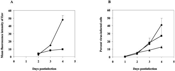
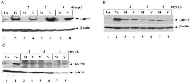
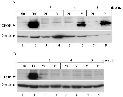
We previously demonstrated that mink cells undergo apoptosis after MCF13 murine leukemia virus (MLV) infection. In this study, we observed that virus-infected mink epithelial cells had significantly larger amounts of steady-state levels of MCF13 MLV envelope precursor protein (gPr80(env)) than did Mus dunni fibroblasts, which are resistant to virus-induced cytopathicity. Infection of mink cells with the noncytopathic NZB-9 MLV did not result in the accumulation of gPr80(env). MCF13 MLV infection of mink cells produced low cell surface expression of envelope glycoprotein and less efficient spread of infectious virus. Western blot analysis of mink epithelial cells infected with MCF13 MLV showed an increase in GRP78/BiP, which was not observed for either mink cells infected with NZB-9 MLV or M. dunni fibroblasts infected with MCF13 MLV. MCF13 MLV infection of mink cells also resulted in a significant upregulation of CHOP/GADD153. These results indicate that the accumulation of MCF13 MLV gPr80(env) triggers endoplasmic reticulum stress, which may mediate apoptosis in mink epithelial cells.


Figures

FIG. 1.
FIG. 1.
Accumulation of MCF13 MLV gPr80env in mink epithelial cells. (A) Western blot analysis of cellular extracts at indicated times p.i. from mink epithelial cells (ATCC CCL64) (lanes 3-5) or M. dunni fibroblasts (ATCC CRL2017) (lanes 7-9) infected with MCF13 MLV at an MOI of 3. M refers to protein extract from mock-infected cells, which were treated only with Polybrene (lanes 2 and 6). Lane 1 (V) consists of an aliquot of purified virions of MCF13 MLV produced by M. dunni fibroblasts. gpPr80env and gp70 bands are indicated by arrowheads. β-Actin was detected as a loading control for cellular extracts. (B) Ratio of gPr80env present in mink cells relative to M. dunni fibroblasts was calculated from densitometry of protein bands in the Western blot in Fig. 1A. (C) Western blot analysis of cell extracts from mink epithelial cells infected with either NZB-9 MLV at an MOI of 3 (lanes 3-5) or mock infected (M, lane 2). (D) Mean ratios of gp70 relative to gPr80env for mink epithelial cells (open bars) and M. dunni fibroblasts (filled bars) are shown. Mean values and standard deviations were calculated from the results of two independent experiments. The differences between mink cells and M. dunni fibroblasts at days 3 and 4 were significant (P = 0.03 using Student's t test).
FIG. 2.
FIG. 2.
Mink epithelial cells have low cell surface expression of MCF13 MLV Env and inefficient virus spread. (A) Mean values and standard deviations of mean fluorescence intensity of Env expression as detected by flow cytometry analysis of mink epithelial cells (▪) and M. dunni fibroblasts (♦) after MCF13 MLV infection over time were calculated from two independent experiments. (B) Percent mink epithelial cells infected with either MCF13 (▴) or NZB-9 MLV (▪) and percent M. dunni fibroblasts infected with MCF13 MLV(♦). Values represent means and standard deviations calculated from the results of two independent experiments.
FIG. 3.
FIG. 3.
MCF13 MLV upregulates GRP78 in mink epithelial cells. Western blot analysis of cellular extracts from (A) mink cells infected with MCF13 MLV (V, lanes 4, 6, and 8) or mock infected (M, lanes 3, 5, and 7). (B) Mink cells infected with NZB-9 MLV (V, lanes 4, 6, and 8) or mock infected (M, lanes 3, 5, and 7). (C) M. dunni fibroblasts infected with MCF13 MLV (V, lanes 4, 6, and 8) or mock infected (M, lanes 3, 5, and 7). Protein extracts from cells exposed to 1 μg of tunicamycin per ml for 24 h (Tu, lanes 2) or untreated (Un, lanes 1) were used for control for the ER stress response. The Western blots are representative of results from at least two independent experiments.
FIG. 4.
FIG. 4.
CHOP induction in mink epithelial cells by MCF13 MLV infection. (A) Western blot analysis of total cellular extracts from mink cells infected with MCF13 MLV (V, lanes 4, 6, and 8) or mock infected (M, lanes 3, 5, and 7). (B) Analysis of mink cells infected with NZB-9 MLV (V, lanes 4, 6, and 8) or mock infected (M, lanes 3, 5, and 7). Cell extracts from mink cells exposed to tunicamycin (Tu, lanes 2) or untreated (Un, lanes 1) were controls for ER stress.

Similar articles

Cited by

References

    1. Bonzon, C., and H. Fan. 1999. Moloney murine leukemia virus-induced preleukemic thymic atrophy and enhanced thymocyte apoptosis correlate with disease pathogenicity. J. Virol. 73:2434-2441. - PMC - PubMed
    1. Chattopadhyay, S. K., M. W. Cloyd, D. L. Linemeyer, M. R. Lander, E. Rands, and D. R. Lowy. 1982. Cellular origin and role of mink cell focus-forming viruses in murine thymic lymphomas. Nature 295:25-31. - PubMed
    1. Dimcheff, D. E., S. Askovic, A. H. Baker, C. Johnson-Fowler, and J. L. Portis. 2003. Endoplasmic reticulum stress is a determinant of retrovirus-induced spongiform neurodegeneration. J. Virol. 77:12617-12629. - PMC - PubMed
    1. Donahue, P. R., S. L. Quackenbush, M. V. Gallo, C. M. deNoronha, J. Overbaugh, E. A. Hoover, and J. I. Mullins. 1991. Viral genetic determinants of T-cell killing and immunodeficiency disease induction by the feline leukemia virus FeLV-FAIDS. J. Virol. 65:4461-4469. - PMC - PubMed
    1. Einfeld, D. 1996. Maturation and assembly of retroviral glycoproteins. Curr. Top. Microbiol. Immunol. 214:133-176. - PubMed

Publication types

MeSH terms