Review
. 2004 Nov;63 Suppl 2(Suppl 2):ii84-ii89.
doi: 10.1136/ard.2004.028316.

Chemokines: role in inflammation and immune surveillance

Affiliations
Review

Chemokines: role in inflammation and immune surveillance

B Moser et al. Ann Rheum Dis. 2004 Nov.

Abstract

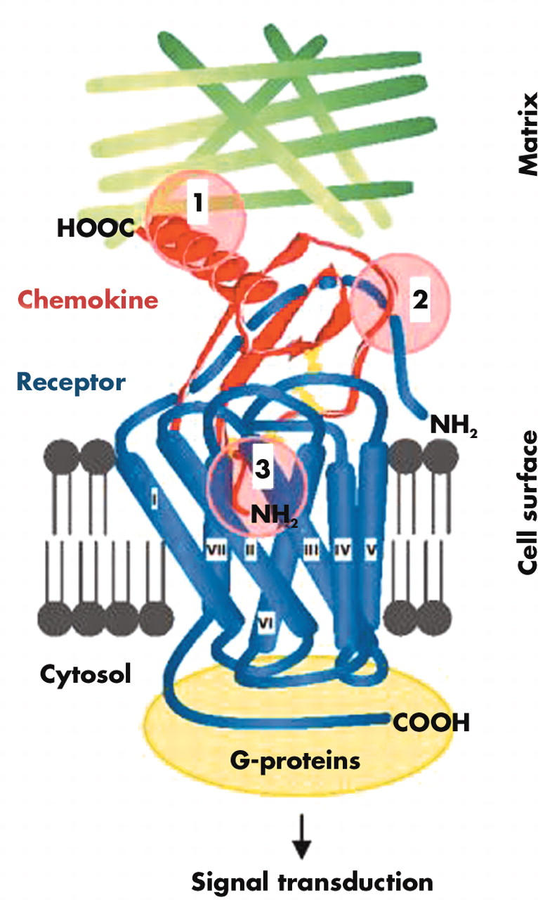
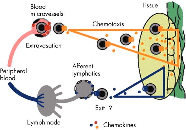
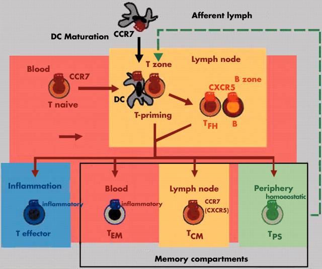
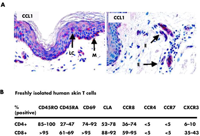
Chemotactic migration of leucocytes largely depends on adhesive interaction with the substratum and recognition of a chemoattractant gradient. Both aspects, cell adhesion and chemotaxis, are regulated by members of the family of chemotactic cytokines (chemokines) comprising structurally related and secreted proteins of 67-127 amino acids in length. Breakdown in the control of leucocyte mobilisation contributes to chronic inflammatory diseases and, hence, interference with chemokine function is a promising approach for the development of novel anti-inflammatory medication. Chemokines target all types of leucocyte, including haematopoietic precursors, mature leucocytes of the innate immune system as well as naive, memory, and effector lymphocytes. The combinatorial diversity in responsiveness to chemokines ensures proper tissue distribution of distinct leucocyte subsets under normal and inflammatory/pathological conditions. Here, we discuss recent views on the role of chemokines in controlling tissue localisation of human memory T cells under steady state (non-inflamed) conditions. Emphasis is placed on a concept describing distinct subsets of memory T cells according to their primary residence in peripheral blood, secondary lymphoid tissues, or peripheral (extralymphoid) tissues.


Figures

Figure 1
Figure 1
Chemokine–receptor interactions. Chemokine receptors are embedded into the membrane by seven transmembrane domains. The NH2-terminus and three extracellular loop regions are involved in chemokine binding, and the COOH-terminus and three intracellular loop regions participate in G-protein mediated signal transduction. The three sites in chemokines that are essential for function include (1) the matrix fixation site in the COOH terminal α-helix or core structure, (2) the N loop region enabling initial receptor contact, and (3) the NH2-terminus, which interacts with the chemokine binding pocket formed by the transmembrane regions of the chemokine receptor.
Figure 2
Figure 2
Recruitment, localisation, and tissue exit of circulating leucocytes. Tissue localisation of leucocytes involves two distinct and sequential processes, termed extravasation and chemotaxis. During extravasation, blood leucocytes interact with adhesion molecules on the luminal side of blood vessels and, upon chemokine receptor triggering, become firmly attached and transmigrate through the epithelial barrier. Subsequent chemotaxis guides the perivascular leucocytes to the cellular source(s) of chemokines, which enables cellular colocalisation and subsequent execution of leucocyte function. Eventually, leucocytes exit the tissue via afferent lymphatic vessels to reach draining lymph nodes (LNs) and peripheral blood.
Figure 3
Figure 3
Chemokine receptors define distinct migratory T cell subsets. Naive T cells develop into effector and memory T cells accompanied by major changes in chemokine receptor expression and responsiveness to chemokines. Effector T cells are short lived and efficiently home to sites of inflammation. Memory T cells, in contrast, are long lived and are classified here according to their principal site of residence. TEM cells mainly reside in peripheral blood and are ready to respond to inflammatory chemokines at sites of immune response. Resting TEM cells lack CCR7 and are excluded from lymph nodes (LNs) and Peyer's patches (PPs). TCM cells express CCR7 and adhesion molecules for homing to T zones of secondary lymphoid tissues. Their principal function is the screening of LNs and PPs for the presence of recall antigens presented by local dendritic cells (DCs). Finally, TPS cells primarily reside in healthy (extralymphoid) peripheral tissues whereas they are rare in peripheral blood, LNs, and PPs. Their primary function is immune surveillance at sites of previous antigen encounter. B, B cells; T naive; naive T cells; T effector, effector T cells, TCM, central memory T cells; TEM, effector memory T cells; TFH, follicular B helper T cells; TPS, peripheral immune surveillance T cells; inflammatory and homoeostatic, receptors for inflammatory and homoeostatic chemokines, respectively.
Figure 4
Figure 4
Complex composition of chemokines and chemokine receptors in rheumatoid arthritis. (A) As an example, inflammatory cells in the affected synovial tissue express Th 1 typical chemokine receptors CXCR3 and CCR5 but not the CCR3, which is more prominent on Th 2 cells. (B) A multitude of chemokines were detected in the synovial fluid and inflamed synovial tissue. The chemokines are listed according to their receptor selectivity and target cells. Of note, the condition of rheumatoid arthritis leads to the generation of all chemokines necessary for recruitment of the full complement of effector cells. BCA, B cell attracting chemokine; ELC, EBI-1-ligand chemokine; ENA, epithelial cell derived neutrophil activating protein; GCP, granulocyte chemotactic protein; GRO, growth related oncogene; IP, interferon α inducible protein; LARC, liver and activation regulated chemokine; MCP, monocyte chemoattractant protein; Mig, monokine induced by interferon γ; MIP, macrophage inflammatory protein; RANTES, regulated on activation, normal T cell expressed and secreted; SDF-1, stromal cell derived factor 1; SLC, secondary lymphoid tissue chemokine.
Figure 5
Figure 5
CCR8 marks skin selective immune surveillance T (TPS) cells. (A) CCL1, the only ligand chemokine for CCR8, is expressed in small amounts by epidermal Langerhans cells (LC) and melanocytes (M) as well as blood vessels in the superficial dermal plexus of normal (non-inflamed) human skin. (B) Summary of phenotypic characteristics of freshly isolated T cells from normal human skin. Please note the abundance of CCR8, which is in striking contrast to the low level of expression of other chemokine receptors that are frequently found on effector/memory T cells in peripheral blood. E, microvascular endothelia.

References

    1. Nat Rev Immunol. 2002 Dec;2(12):982-7 - PubMed
    1. J Immunol. 2002 Nov 15;169(10):5838-43 - PubMed
    1. J Exp Med. 2003 Jun 2;197(11):1537-49 - PubMed
    1. Trends Immunol. 2003 Jun;24(6):327-34 - PubMed
    1. Blood. 2003 Aug 1;102(3):789-94 - PubMed

Publication types

MeSH terms