. 2005 Jan;16(1):153-61.
doi: 10.1091/mbc.e04-05-0413. Epub 2004 Oct 13.

Role of Unc104/KIF1-related motor proteins in mitochondrial transport in Neurospora crassa

Affiliations

Role of Unc104/KIF1-related motor proteins in mitochondrial transport in Neurospora crassa

Florian Fuchs et al. Mol Biol Cell. 2005 Jan.

Abstract

Eukaryotic cells use diverse cytoskeleton-dependent machineries to control inheritance and intracellular positioning of mitochondria. In particular, microtubules play a major role in mitochondrial motility in the filamentous fungus Neurospora crassa and in mammalian cells. We examined the role of two novel Unc104/KIF1-related members of the kinesin family, Nkin2 and Nkin3, in mitochondrial motility in Neurospora. The Nkin2 protein is required for mitochondrial interactions with microtubules in vitro. Mutant hyphae lacking Nkin2 show mitochondrial motility defects in vivo early after germination of conidiospores. Nkin3, a member of a unique fungal-specific subgroup of small Unc104/KIF1-related proteins, is not associated with mitochondria in wild-type cells. However, it is highly expressed and recruited to mitochondria in Deltankin-2 mutants. Mitochondria lacking Nkin2 require Nkin3 for binding to microtubules in vitro, and mitochondrial motility defects in Deltankin-2 mutants disappear with up-regulation of Nkin3 in vivo. We propose that mitochondrial transport is mediated by Nkin2 in Neurospora, and organelle motility defects in Deltankin-2 mutants are rescued by Nkin3. Apparently, a highly versatile complement of organelle motors allows the cell to efficiently respond to exogenous challenges, a process that might also account for the great variety of different mitochondrial transport systems that have evolved in eukaryotic cells.


Figures
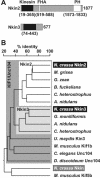
Figure 1.
Figure 1.
Nkin2 and Nkin3 are novel members of the Unc104/KIF1 subfamily of kinesin-related motor proteins. (A) Domain structure of Nkin2 and Nkin3. Homologies to known protein domains were revealed by searching the Pfam database (Bateman et al., 2002). Kinesin motor domains are depicted in black, FHA domains in light gray, and PH domains in white. Numbers indicate amino acid residues defining the borders of the predicted domains, and the C termini of the proteins, respectively. Protein domains are drawn to scale. (B) Homology tree of representative kinesin family members. Fungal homologues sharing the same domain structure with Nkin2 are highlighted by a white box. This group includes predicted proteins from Magnaporthe grisea (genome annotation number MG09255.4), Gibberella zeae (FG10189.1), Botryotinia fuckeliana (gene KLP8), Cochliobolus heterostrophus (KLP8), and Aspergillus nidulans (AN7547.2). Fungal homologues closely resembling Nkin3 are highlighted by a light gray box. This group includes predicted proteins from Gibberella moniliformis (KLP7), A. nidulans (AN6863.2), and Cochliobolus heterostrophus (KLP7). Other members of the KIF1/Unc104 family sharing the same domain structure with Nkin2 are Kin3 of Ustilago maydis (Wedlich-Söldner et al., 2002), KIF1B of Mus musculus (Nangaku et al., 1994), Unc104 of C. elegans (Otsuka et al., 1991), and Unc104 of D. discoideum (Pollock et al., 1999). Nkin (Steinberg and Schliwa, 1995) and Kif5b (Tanaka et al., 1998) are conventional kinesins. The tree was constructed using DNAMAN software (Lynnon BioSoft, Vaudreuil, Canada).
Figure 2.
Figure 2.
Nkin2 is peripherally associated with the mitochondrial outer membrane. (A) Subfractionation of cells. Wild-type cells were fractionated by differential centrifugation into PMS (lane 1) and a crude mitochondrial fraction (lane 2). Crude mitochondria were either further purified by sucrose gradient centrifugation (lane 3) or used for preparation of OMVs (lane 4). Fifty micrograms of protein of each fraction was analyzed by SDS-PAGE and immunoblotting by using specific antisera against Nkin2, Nkin3, and Nkin. The integral outer membrane protein, Tom40, served as a marker for mitochondria, and the peroxisomal protein, Pex14, served as a marker for nonmitochondrial membranes. (B) Salt-extraction of mitochondria. Isolated mitochondria were left untreated on ice (lane 1) or extracted with 1 M KCl and reisolated by centrifugation (lane 2). Proteins were precipitated from the salt extract with TCA (lane 3). Fifty micrograms of protein of each fraction was analyzed by SDS-PAGE and immunoblotting. Tom40 served as a marker for the mitochondrial outer membrane, and CCHL, a soluble protein of the intermembrane space, was used to control for integrity of the outer membrane.
Figure 3.
Figure 3.
Antibodies against Nkin2 inhibit mitochondrial binding to microtubules in vitro. (A) Flotation assay. Isolated mitochondria were preincubated in the absence or presence of the antibodies indicated on the right-hand side. Then, mitochondria were reisolated by centrifugation, incubated with taxol-stabilized microtubules in the absence of ATP and floated in a sucrose density gradient. Fractions were harvested from the gradient, and proteins were precipitated and analyzed by SDS-PAGE and immunoblotting by using antibodies against the proteins indicated on the left-hand side. Only fractions 1 (containing floated mitochondria as controlled by blotting against porin), 3 (from the middle of the gradient), and 5 (containing nonfloated proteins) are shown. P, pellet from the bottom of the gradient. (B) Visual assay. Mitochondria were isolated from a Neurospora strain expressing mtGFP and preincubated in the absence or presence of the indicated antibodies. Then, rhodamine-labeled microtubules (rho-MT) were added, incubation was continued in the absence of ATP, and samples were analyzed by fluorescence microscopy. Bar, 5 μm.
Figure 4.
Figure 4.
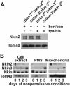
Nkin3 is up-regulated and recruited to mitochondria in the absence of Nkin2 in heterokaryotic cells. (A) Depletion of Nkin2 in heterokaryotic cells. The HP1 wild-type strain (nkin-2WT/nkin- 2WT) and a heterokaryotic mutant strain (Δnkin-2/nkin-2WT) were grown for 3 d in liquid minimal medium supplemented either with benomyl and pantothenic acid (ben/pan) or p-fluoro-DL-phenylalanine and histidine (fpa/his). Equal amounts of cell extracts were analyzed by SDS-PAGE and immunoblotting. (B) Up-regulation and mitochondrial association of Nkin3 upon depletion of Nkin2. Heterokaryotic Δnkin-2/nkin-2WT cells were grown for the indicated time periods in liquid minimal medium supplemented with fpa/his. Total cell extracts, PMSs, and mitochondria were prepared, and equal amounts of protein were analyzed by SDS-PAGE and immunoblotting.
Figure 5.
Figure 5.
Nkin3 is up-regulated and binds to mitochondria in growing homokaryotic cells lacking Nkin2. (A) Up-regulation and mitochondrial association of Nkin3 in homokaryotic Δnkin-2 mutants. The HP1 wild-type strain (nkin-2WT/nkin-2WT), a heterokaryotic mutant (Δnkin-2/nkin-2WT), and two independently isolated homokaryotic mutants (Δnkin-2) were grown for 16 h in fpa/his-containing medium. Then, cells were analyzed as in Figure 4B. (B) Growth of Δnkin-2 cells. Wild-type (WT) and Δnkin-2 cells were inoculated in the middle of a petri dish containing Vogel's minimal medium agar and grown over night at 25°C. Bar, 1 cm. (C) Mitochondrial association of Nkin3 in Δnkin-2 cells. Δnkin-2 cells were subfractionated and analyzed as in Figure 2A. (D) Nkin3 level in conidiospores. Cell extracts were prepared from conidiospores of the homokaryotic Δnkin-2 strain and its isogenic wild-type (WT) and analyzed by SDS-PAGE and immunoblotting. (E) Up-regulation of Nkin3 in newly germinated Δnkin-2 cells. Conidiospores of the homokaryotic Δnkin-2 strain were allowed to germinate and grow for the indicated time periods. Then, total cell extracts were prepared and analyzed by SDS-PAGE and immunoblotting. For comparison, a cell extract of a 14 h old wild-type culture is shown.
Figure 6.
Figure 6.
Antibodies against Nkin3 inhibit binding of Δnkin-2 mitochondria to microtubules in vitro. Mitochondria were isolated from a Δnkin-2 mutant and subjected to the microtubule flotation assay as in Figure 3A.
Figure 7.
Figure 7.
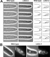
Cells lacking Nkin2 show altered mitochondrial motility and morphology. (A) Wild-type and Δnkin-2 conidiospores were allowed to germinate and grow over night on medium-covered microscope slides. Then, mitochondrial behavior in growing hyphae was analyzed by computer-enhanced phase contrast videomicroscopy (2 images per second). The complete datasets are available in two movies covering 30 s of hyphal growth (see Supplemental Movie 1 for behavior of wild-type mitochondria and movie 2 for Δnkin-2 mitochondria). They are representative examples of 10 independent experiments. Differences in cellular growth rate are due to variations between individual cells. Right, representative individual organelles near the hyphal tip have been highlighted. Bar, 5 μm. (B) Wild-type and Δnkin-2 cells were grown for 7 h after germination of conidiospores, stained with 10 μg/ml rhodamine B hexyl ester, and analyzed by phase contrast (left) and fluorescence (right) microscopy. Bar, 5 μm.

Similar articles

Cited by

References

    1. Bateman, A., et al. (2002). The Pfam protein families database. Nucleic Acids Res. 30, 276-280. - PMC - PubMed
    1. Bereiter-Hahn, J. (1990). Behavior of mitochondria in the living cell. Int. Rev. Cytol. 122, 1-63. - PubMed
    1. Boldogh, I. R., Yang, H.-C., Nowakowski, W. D., Karmon, S. L., Hays, L. G., Yates III, J. R., and Pon, L. A. (2001a). Arp2/3 complex and actin dynamics are required for actin-based mitochondrial motility in yeast. Proc. Natl. Acad. Sci. USA 98, 3162-3167. - PMC - PubMed
    1. Boldogh, I. R., Yang, H.-C., and Pon, L. A. (2001b). Mitochondrial inheritance in budding yeast. Traffic 2, 368-374. - PubMed
    1. Borkovich, K. A., et al. (2004). Lessons from the genome sequence of Neurospora crassa: tracing the path from genomic blueprint to multicellular organism. Microbiol. Mol. Biol. Rev. 68, 1-108. - PMC - PubMed

Publication types

MeSH terms

Substances