. 2004 Oct 13;24(41):8940-9.
doi: 10.1523/JNEUROSCI.3065-04.2004.

Cortical GABA interneurons in neurovascular coupling: relays for subcortical vasoactive pathways

Affiliations

Cortical GABA interneurons in neurovascular coupling: relays for subcortical vasoactive pathways

Bruno Cauli et al. J Neurosci. .

Abstract

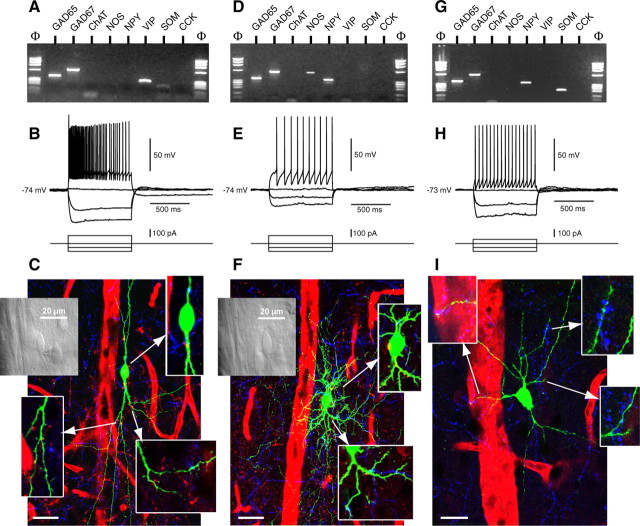
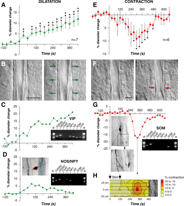
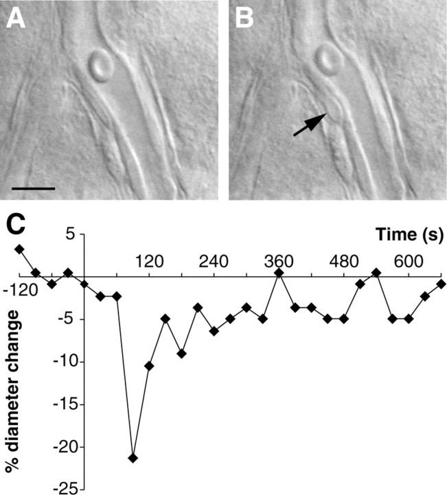
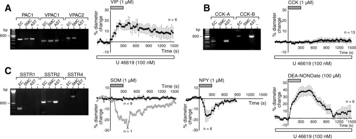
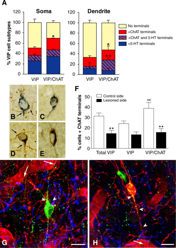
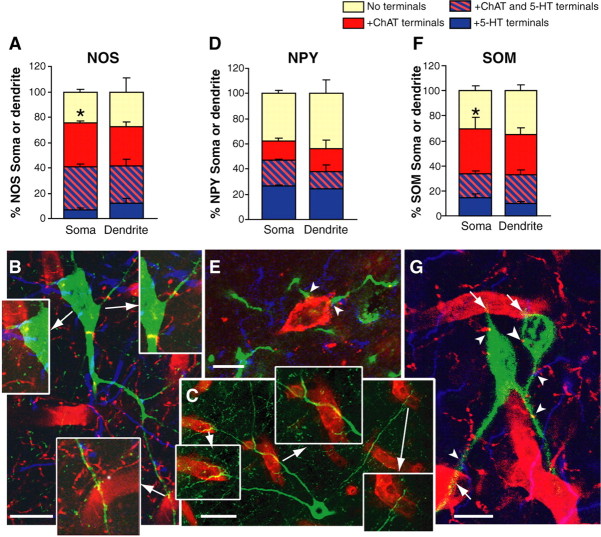
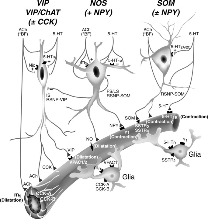
The role of interneurons in neurovascular coupling was investigated by patch-clamp recordings in acute rat cortical slices, followed by single-cell reverse transcriptase-multiplex PCR (RT-mPCR) and confocal observation of biocytin-filled neurons, laminin-stained microvessels, and immunodetection of their afferents by vasoactive subcortical cholinergic (ACh) and serotonergic (5-HT) pathways. The evoked firing of single interneurons in whole-cell recordings was sufficient to either dilate or constrict neighboring microvessels. Identification of vasomotor interneurons by single-cell RT-mPCR revealed expression of vasoactive intestinal peptide (VIP) or nitric oxide synthase (NOS) in interneurons inducing dilatation and somatostatin (SOM) in those eliciting contraction. Constrictions appeared spatially restricted, maximal at the level of neurite apposition, and were associated with contraction of surrounding smooth muscle cells, providing the first evidence for neural regulation of vascular sphincters. Direct perfusion of VIP and NO donor onto the slices dilated microvessels, whereas neuropeptide Y (NPY) and SOM induced vasoconstriction. RT-PCR analyses revealed expression of specific subtypes of neuropeptide receptors in smooth muscle cells from intracortical microvessels, compatible with the vasomotor responses they elicited. By triple and quadruple immunofluorescence, the identified vasomotor interneurons established contacts with local microvessels and received, albeit to a different extent depending on interneuron subtypes, somatic and dendritic afferents from ACh and 5-HT pathways. Our results demonstrate the ability of specific subsets of cortical GABA interneurons to transmute neuronal signals into vascular responses and further suggest that they could act as local integrators of neurovascular coupling for subcortical vasoactive pathways.


Figures

Figure 1.
Figure 1.
Molecular, electrophysiological, and morphological characterizations of perivascular neurons. A, Molecular analysis of a perivascular neuron expressing GAD65, GAD67, and VIP. B, Firing pattern of the same interneuron. Application of a 100 pA depolarizing current pulse induced a discharge of action potentials with a marked frequency adaptation (top trace). Note the augmentation after a marked reduction of spike amplitude. C, Visualization of the same neuron immunodetected for biocytin (green) revealed its bipolar-bitufted morphology, projections to local blood vessels (red), and numerous ACh (red) and 5-HT (blue) afferents. Top left, Inset, Morphology of the neuron as seen under infrared illumination. Insets, ACh (red) and 5-HT (blue) varicosities contacting the neuron soma and dendrites (green) in single-plane sections. D, Molecular analysis of another perivascular neuron showing expression of GAD65, GAD67, NOS, and NPY. E, Firing pattern of the same interneuron. Application of a 100 pA depolarizing current induced a discharge of action potentials with a slight increase in firing rate (top trace). Note the delay of the discharge of the first action potential. F, Immunodetection of biocytin (green) in the same neuron revealed a neurogliaform cell with extensive dendritic arborization and appositions on the adjacent blood vessel. Left inset, Morphology of the neuron as seen under infrared illumination. Insets, Somatic ACh (red) and dendritic ACh and 5-HT (blue) contacts seen in single-plane sections. G, Molecular analysis of another perivascular interneuron expressing GAD65, GAD67, NPY, and SOM. H, Firing pattern of the same neuron. Application of a 50 pA depolarizing current induced a discharge of action potentials with no frequency adaptation (top trace). I, The same neuron visualized with biocytin (green) exhibited a triangular morphology with projections to the adjacent blood vessel (left inset). Right insets, Appositions of 5-HT immunostained (blue) varicosities (blue) on the dendrites of the SOM neuron. Arrows in C, F, and I point to insets of the corresponding areas from each neuron. Scalebars: C, F, 30 μm; I, 20 μm.
Figure 2.
Figure 2.
Single interneuron stimulation induces dilation or constriction of cortical blood vessels. A, C, D, E, G, Onsets of evoked firing start at a time of 0 sec. A, Mean values ± SEM of dilating responses induced by evoked firing of different cortical interneurons (n = 7). Dilatation develops in phase with electrical stimulation. *p < 0.05 and **p < 0.01 from prestimulation value. B, F, Images of blood vessels before (left) and after (right) electrical stimulation (120 sec) of the interneurons shown in C and G, respectively. Scale bars, 10 μm. The arrows indicate regions of high vascular reactivity. C, Temporal response of the blood vessel shown in B. Inset, Molecular characterization of the stimulated interneuron showing expression of GAD65, GAD67, NPY, VIP, SOM, and CCK. D, Temporal response of a blood vessel evoked by electrical stimulation (30 sec) of another interneuron. Note the reversibility of the dilatation. Left inset, Morphology of the stimulated neuron (biocytin revealed with DAB; brown) showing a right horizontal dendritic arborization coursing toward the responsive blood vessel (immunodetected with laminin and the SG reagent; blue gray). Right inset, Molecular characterization of the stimulated interneuron showing expression of GAD65, GAD67, NOS, and NPY. E, Mean values ± SEM of contracting responses evoked by electrical stimulation of different cortical interneurons (n = 6; *p < 0.05 from prestimulation value). G, Temporal response of the blood vessel shown in F. Note the delay and the reversibility of the contraction. Left inset, Morphology of the stimulated neuron immunodetected for biocytin (DAB) showed a vertically oriented dendritic arborization. Bottom image shows a dendritic branch projecting toward the responsive blood vessel at the level of the highest vascular reactivity (red arrow in F; right panel). Right inset, Molecular characterization of the stimulated interneuron showing expression of GAD65, GAD67, and SOM. H, Spatiotemporal response of the blood vessel shown in F disclosing a delayed and spatially restricted contraction (hot spot; red). Amplitude of contraction is color coded (scale in percentage of contraction). The arrows indicate the duration of evoked firing.
Figure 3.
Figure 3.
Contractile element in a cortical microvessel. Image of a small blood vessel showing the diameter and smooth muscle cell of the microvessel before (A) and after (B) the evoked firing (120 sec) of a SOM interneuron identified by single-cell RT-PCR. The arrow in B shows the contraction of a smooth muscle cell during the stimulation. Note the reduction of luminal diameter. C, Temporal response of the same blood vessel, onset of evoked firing started at a time of 0 sec and lasted 120 sec. Note the reversibility of the contraction. Scale bars, 10 μm.
Figure 4.
Figure 4.
Molecular expression and vasomotor effects of microvascular receptors. Gel electrophoresis of PCR products for VIP (A), CCK (B), and SOM (C) receptor subtypes expressed in cultures of EC, SMC, and AST. Only products expressed in 50% or more of the cell cultures are illustrated. EC expressed all subtypes of VIP and CCK receptors and only SSTR1 and SSTR2. SMC were found to express all VIP receptor subtypes, no CCK receptors, and SSTR2 and SSTR4 receptors. In contrast, AST showed expression of PAC1, VPAC1, CCK-A, and CCK-B receptors and SSTR2 receptor subtypes. Samples without reverse transcriptase (-) were included to control for possible contamination. Vascular responses (mean ± SEM) were evoked in cortical slices by bath application of different substances known to colocalize in different subsets of GABA interneurons. After preconstriction with U46619, VIP induced a dilatation in 6 of 10 microvessels (A, right panel), whereas CCK failed to elicit any vasomotor response (B, right panel; n = 13). At baseline, most microvessels tested (9 of 10) were unresponsive to bath application of SOM (C, second panel) except for one, which constricted in phase with the peptide application (open circles). A majority (6 of 7) of vessels reversibly constricted after NPY application, whereas the NO donor DEA NONOate strongly and reversibly dilated microvessels (n = 6 of 14; C, third and forth panels, respectively).
Figure 5.
Figure 5.
Identification of subsets of VIP interneurons and their ACh and 5-HT afferents. A, In triple-labeled sections for VIP, ChAT, and 5-HT, comparable proportions of soma (∼40%) and dendrites (∼20%) from VIP and VIP/ChAT neurons received 5-HT afferents (blue box). In contrast, a larger proportion of VIP/ChAT than VIP neurons was contacted by ACh afferents (red box) on both cell soma (n = 216; p < 0.05) and dendrites (n = 186; p < 0.05), and a larger proportion of VIP/ChAT dendrites also received both types of afferents (**p < 0.01; hatched red and blue box) compared with VIP dendrites. B-F, Effect of unilateral basal forebrain lesion on the ACh afferents to VIP interneurons as evaluated in semithin sections labeled for VIP (SG kit; blue) and ChAT (DAB; brown). Quantitative analysis of VIP and VIP/ChAT neurons on the control (B, D; n = 476 cells) and lesioned (C, E; n = 496 cells) sides, respectively, showed a significant loss of ACh input after lesion of substantia innominata (F). Approximately 50% of the ACh innervation to the total VIP cell population (**p < 0.01) originated in the basal forebrain, with VIP/ChAT cells being significantly denervated (60%; **p < 0.01) but not the VIP (44%; NS) cells. Also, the analysis on the control side confirmed that VIP/ChAT cells received more ACh afferents than VIP cells (∝; p < 0.05). G, Quadruple immunofluorescence of a perivascular VIP neuron (green; Cy2) contacting a neighboring blood vessel (arrows; laminin-immunodetected with Cy3, red) and receiving both ACh (ChAT and Cy3; red) and 5-HT (5-HT and Cy5; blue) afferent terminals on its cell body or proximal dendrites (arrowheads). H, Quadruple immunofluorescence as in G but showing a VIP/ChAT interneuron (red/yellow) in contact with a blood vessel (arrow) and receiving dual ACh (red) and 5-HT (blue) innervations (arrowheads). Scale bars, 10 μm.
Figure 6.
Figure 6.
NOS, NPY, and SOM interneurons and their ACh (red box), 5-HT (blue box), and dual ChAT and 5-HT (hatched red and blue box) afferent inputs. A, Quantitative analysis of these innervations in NOS neurons (n = 90 somata and 85 dendrites) showed that a larger proportion of cell bodies was contacted by ACh varicosities compared with 5-HT afferents (*p < 0.05). Note also that a large proportion (∼30%) of NOS neurons received both types of innervations. B, Quadruple immunofluorescence (NOS, ChAT, 5-HT, and laminin) showing a NOS interneuron (green) projecting to a surrounding blood vessel (bottom inset) and receiving both ACh (red) and 5-HT (blue) afferents in single-plane sections. Insets, ACh and 5-HT contacts are abundant on both cell soma and dendrites. C, Double immunostaining for NOS (green) and laminin (red) depicting the extensive perivascular projections from a single NOS neuron with nearby and remotely located microvessels (insets). D, Quantitative analysis of ACh and 5-HT afferents to small NPY neurons that do not colocalize NOS (n = 98 somata and 54 dendrites). Overall, these NPY neurons received comparable ACh (∼32%, ChAT and Cy5; blue) or 5-HT (43%, serotonin transporter and Cy2; green) afferents (shown in E), with 15-19% being contacted by both types of varicosities. F, Quantitative analysis of ACh and 5-HT innervations of SOM neurons (n = 156 somata and 112 dendrites) showed that a larger proportion of these cells (∼60%; *p < 0.05) received ACh terminals on their cell soma compared with 5-HT varicosities, with ∼20% of the cells receiving both types of afferents. G, Two perivascular SOM neurons (Cy2; green) contacting local blood vessels (arrows, laminin and Cy3; red) and receiving both ACh (ChAT and Cy3; red) and 5-HT (5-HT and Cy5; blue) varicosities (arrowheads). Scale bars: E, G, 10 μm; B, 20 μm; C, 30 μm.
Figure 7.
Figure 7.
Schematic representation of the suggested role of cortical interneurons in neurovascular coupling and as possible relay neurons for vasoactive basal forebrain (BF) ACh and brainstem 5-HT systems. Their direct vasomotor effects are thought to be mediated by m5 muscarinic ACh receptor (dilatation) or 5-HT1B receptor (constriction). However, the ability of these systems to target cortical GABA interneurons projecting to local microvessels that are endowed with subtype-specific receptors for vasoactive neuropeptides (VIP, NPY, or SOM) colocalized with GABA strongly support a role of cortical interneurons in modulating microvascular tone. A role for perivascular astrocytes cannot be excluded because they are neuropeptide receptive and are known to release vasodilatory mediators. Note that there is no receptor for CCK on smooth muscle cells and that this peptide failed to induce any vasomotor response. Also, cortical interneurons could serve as local relays in neurovascular coupling for other vasoactive afferent pathways (e.g., glutamate) (see Discussion).

References

    1. Abounader R, Hamel E (1997) Associations between Neuropeptide Y nerve terminals and intraparenchymal microvessels in rat and human cerebral cortex. J Comp Neurol 388: 444-453. - PubMed
    1. Abounader R, Elhusseiny A, Cohen Z, Olivier A, Stanimirovic D, Quirion R, Hamel E (1999) Expression of neuropeptide Y receptors mRNA and protein in human brain vessels and cerebromicrovascular cells in culture. J Cereb Blood Flow Metab 19: 155-163. - PubMed
    1. Bao L, Kopp J, Zhang X, Xu Z-QD, Zhang L-F, Wong H, Walsh J, Hokfelt T (1997) Localization of neuropeptide Y Y1 receptors in cerebral blood vessels. Proc Natl Acad Sci USA 94: 12661-12666. - PMC - PubMed
    1. Bayraktar T, Staiger JF, Acsady L, Cozzari C, Freund TF, Zilles K (1997) Co-localization of vasoactive intestinal polypeptide, gamma-aminobutyric acid and choline acetyltransferase in neocortical interneurons of the adult rat. Brain Res 757: 209-217. - PubMed
    1. Cauli B, Audinat E, Lambolez B, Angulo MC, Ropert N, Tsuzuki K, Hestrin S, Rossier J (1997) Molecular and physiological diversity of cortical nonpyramidal cells. J Neurosci 17: 3894-3906. - PMC - PubMed

Publication types

MeSH terms

LinkOut - more resources