. 2004 Oct 13;24(41):8994-9004.
doi: 10.1523/JNEUROSCI.3184-04.2004.

Alpha2-chimaerin, cyclin-dependent Kinase 5/p35, and its target collapsin response mediator protein-2 are essential components in semaphorin 3A-induced growth-cone collapse

Affiliations

Alpha2-chimaerin, cyclin-dependent Kinase 5/p35, and its target collapsin response mediator protein-2 are essential components in semaphorin 3A-induced growth-cone collapse

Matthew Brown et al. J Neurosci. .

Abstract

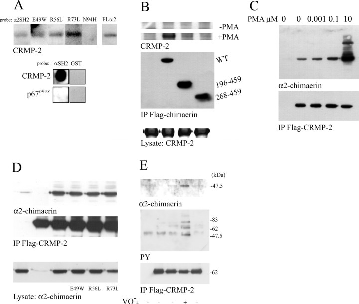
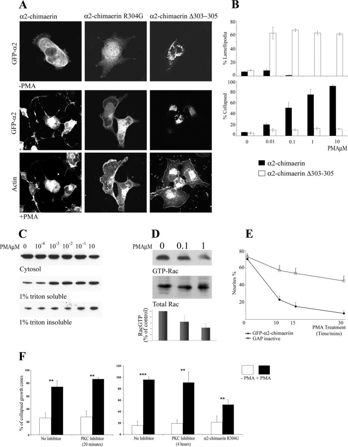
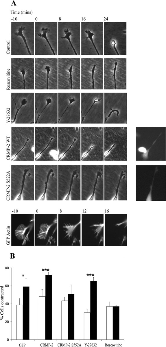
Neurite outgrowth is influenced by positive and negative signals that include the semaphorins, an important family of axonal outgrowth inhibitors. Here we report that the Rac GTPase activating protein (GAP)alpha2-chimaerin is involved in Semaphorin 3A (Sema 3A) signaling. In dorsal root ganglion neurons, Sema 3A-induced growth cone collapse was inhibited by alpha2-chimaerin mutated to eliminate GAP activity or interaction with phosphotyrosine. Activation of alpha2-chimaerin by phorbol ester caused growth cone collapse. Active alpha2-chimaerin interacts with collapsin response mediator protein-2 (CRMP-2) and cyclin-dependent kinase (Cdk) 5/p35 kinase through its SH2 and GAP domains, respectively. Cdk5 phosphorylates CRMP-2 at serine 522, possibly facilitating phosphorylation of serine 518 and threonine 514 by glycogen synthase kinase 3beta (GSK3beta), a kinase previously implicated in Sema 3A signaling. Phosphorylation of CRMP-2 serine 522 was essential for Sema 3A-induced growth cone collapse, which is dependent on Cdk5 but not Rho kinase activity. alpha2-chimaerin, like CRMP-2, can associate with the Sema 3A receptor. These results indicate that active alpha2-chimaerin Rac GAP, Cdk5/p35, and its substrate CRMP-2, are implicated in the dynamics of growth cone guidance initiated through Sema 3A signaling.


Figures

Figure 1.
Figure 1.
Purification of CRMP-2 from rat brain extracts. A, Rat brain extracts (see Materials and Methods) fractionated on S-Sepharose were analyzed by Western blot and overlay asay with 32P-labeled α2-chimaerin SH2 (lanes 1-3) or GAP domain (lanes 4-5) (left). A 64 kDa protein was purified by sequential chromatography of brain soluble fraction (lane 7) on S-Sepharose (lanes 8-11), Q-Sepharose (lanes 12-15), Zinc chelating Sepharose (lanes 16-19), and phenyl Sepharose (lanes 21-23) (Materials and Materials) (Hall et al., 1993). Fractions were eluted from S-Sepharose in a salt gradient, and 0.2 m NaCl fraction (lane 10) was diluted and fractionated on Q-Sepharose. P64 eluted with 0.2 m NaCl (lane 13) and applied to a Zinc chelating column, eluted at pH 6.0 (lane 17) or with 50 mm imidazole (lane 19). The latter, adjusted to 0.4 m, was bound to phenyl-Sepharose and eluted with 0.2 m NH4S04. Eluted fractions were analyzed by overlay assay for α2-chimaerin interaction. Fractions shown are as follows: 0.1 m NaCl, lanes 2, 5, 9, 12; 0.2 m NaCl, lanes 3, 6, 10, 13; 0.3 m NaCl, lanes 11 and 14; 0.4 m NaCl, lane 15; Zn chelate unbound, lane 16; pH 6.0 elution, lane 17; 20 mm imidazole, lane 18; 50 mm imidazole, lane 19; phenyl-Sepharose unbound, lane 20; 0.2 m NH4SO4, lane 21. The final fraction was analyzed by 2-D gel electrophoresis, stained with Coomassie blue, and two p64 spots identified by overlay were cut out for peptide sequence analysis. The peptide sequences that were obtained are underlined in B. B, CRMP-2 sequence. Peptides isolated and sequenced are underlined. C, Western analysis of CRMP-2 and α2-chimaerin in embryonic and neonatal rat brain.
Figure 2.
Figure 2.
CRMP-2 interacts with α2-chimaerin SH2 domain. A, In vitro association of recombinant CRMP-2 with α2-chimaerin. Top, Western blot of recombinant CRMP-2 probed with 32P-labeled SH2 domain, SH2 domain mutants E49W, R56L, R73L, N94H, and full-length α2-chimaerin (FL). Bottom, Dot blot of recombinant CRMP-2 and p67phox control protein, probed with 32P-labeled α2-chimaerin SH2 domain or GST control. B, In vivo interaction of CRMP-2 with α2-chimaerin. Full-length Flag-α2-chimaerin (WT) and N-terminal truncations Flag-α2-chimaerin(196 - 459) and Flag-α2-chimaerin (268 - 459) were coexpressed in COS-7 cells with HA-CRMP-2 in the presence and absence of PMA (10 μm). Western blots of immunoprecipitated (IP) Flag-chimaerin were probed with anti-HA antibody to detect associated CRMP-2. C, Western blots of HA-α2-chimaerin associated with immunoprecipitated Flag-CRMP-2 after coexpression in COS-7 cells treated with PMA 0-10 μm as specified. D, Western blots of α2-chimaerin, α2-chimaerin E49W, α2-chimaerin R56L, and α2-chimaerin R73L immunoprecipitated with Flag-CRMP-2 after coexpression in COS-7 cells treated with PMA (10 μm). E, Western blots of α2-chimaerin (top), phosphotyrosine-containing proteins (detected with RC20 antibody; middle) immunoprecipitated with Flag-CRMP-2 (bottom) from cells of a permanent α2-chimaerin N1E-115 cell line transiently transfected with Flag-CRMP-2.
Figure 3.
Figure 3.
Cdk5 and its neuronal regulator p35 interact with α2-chimaerin GAP domain. A, In vivo interaction of p35 with full-length Flag-α2-chimaerin (WT) and N-terminal truncations Flag-α2-chimaerin (196 - 459) and Flag-α2-chimaerin (268 - 459) immunoprecipitated after their coexpression in COS-7 cells with p35. B, In vivo interaction of Cdk5 with full-length Flag-α2-chimaerin (WT) and N-terminal truncations Flag-α2-chimaerin (39 - 459), Flag-α2-chimaerin (196 - 459), and Flag-α2-chimaerin (268 - 459) immunoprecipitated after coexpression in COS-7 cells with Cdk5. C, Immunoprecipitation of Flag-p35 from COS-7 cells after coexpression with the indicated combinations of Cdk5, kinase inactive Cdk5 N144, and/or GFP-α2-chimaerin. Western blots of α2-chimaerin, Cdk5, and Flag p35 in immunoprecipitates are shown. D, Coimmunoprecipitation of α2-chimaerin with Flag-p35 after coexpression in COS-7 cells treated with increasing amounts of PMA (0-10 μm) as indicated. E, Coimmunoprecipitation of Cdk5 with Flag-α2-chimaerin after coexpression in COS-7 cells treated with PMA (0-10 μm) as indicated. F, Coimmunoprecipitation with anti-Flag-p35 of Cdk5 andα2-chimaerin from COS-7 cells expressing all three proteins and treated with PMA (0-10 μm) as indicated. G, Immunoprecipitation with anti-Flag-p35 of GFP-α2-chimaerin (WT), α2-chimaerin N94H, α2-chimaerin R304G, and α2-chimaerin Δ303-305 after coexpression in COS-7 cells.
Figure 4.
Figure 4.
Phorbol ester activation and membrane translocation of α2-chimaerin, autoinhibited in cytosol. A, PMA activation of GFP-α2-chimaerin expressed in COS-7 cells (A, B) and of α2-chimaerin in N1E-115 cell lines (C-E). A, Cell morphology and distribution of α2-chimaerin, α2-chimaerin R304G, and α2-chimaerin Δ303-305 (2 GAP in active mutants) (top) in the presence and absence of PMA treatment. Phalloidin-stained actin is shown for the PMA-treated cells (bottom). B, Quantitation of COS-7 cells showing ruffling (lamellipodia; top) and collapse (bottom) in response to increasing concentrations of PMA after transfection with α2-chimaerin (filled columns) or GAP inactive α2-chimaerin Δ303-305 (open columns). C, Western analysis of α2-chimaerin from permanent α2-chimaerin N1E-115 cell line treated with increasing concentrations of PMA and fractionated into soluble, membrane, and 1% Triton-insoluble fraction (Kozma et al., 1996). D, Rac activity, determined by Rac-GTP pull-down with GST PAK GTPase binding region, in α2-chimaerin permanent cells treated with PMA at the indicated concentrations; Western blot with anti-Rac antibody shows total Rac and Rac-GTP bound to a GST-PAK CRIB (Cdc42/Rac interactive binding) affinity matrix. E, Effect of PMA treatment on neurites of N1E-115 cells, differentiated by serum starvation and transiently transfected with GFP-α2-chimaerin or GFP-α2-chimaerin GAP inactive mutant. Results show the average of three experiments (SEM). F, DRG neurons treated with PMA 100 nm (filled columns) for 30 min, in the presence or absence PKC inhibitor bisindolylmaleimide (10 μm) after inhibitor pretreatment of 20 min or 4 hr, or after transfection with GAP mutant GFP-α2-chimaerin R304G (see Materials and Methods). Cells were fixed and stained with phalloidin tetramethylrhodamine isothiocyanate, and GFP-transfected cells with collapsed growth cones were quantitated. PMA-induced collapse of α2-chimaerin R304G-transfected cells was significantly less than control cells plus or minus PKC inhibition; p > 0.008 and p > 0.003, respectively.
Figure 8.
Figure 8.
α2-chimaerin is involved in growth cone collapse in DRG neurons. A, DRG neurons in culture stained with α2-chimaerin antibody and with Cy5-conjugated anti-rabbit IgG secondary antibody (red) and phalloidin-FITC (green). B, DRG neurons transfected with GFP, GFP α2-chimaerin, GFP-α2-chimaerin R304G, and α2-chimaerin R73L were treated plus or minus Sema 3A as described in Materials and Methods, and collapsed growth cones were quantified. **p < 0.01; ***p < 0.001; growth cone collapse significantly different in presence of either PMA (B) or Sema 3A (C) than in its absence. C, DRG neurons were transfected with GFP, GFP plus ArhGAP15, or GFP plus ArhGAP15 GAP inactive. Neurons were fixed and stained for actin, and collapsed growth cones were quantified. All three demonstrated an equally significant increase in Sema 3A-induced growth cone collapse.
Figure 5.
Figure 5.
CRMP-2 serine 522 is a novel substrate of Cdk5 and required for phosphorylation by GSK3 β. A,B, Flag-CRMP-2 (WT) and mutants of the sites indicated were synthesized in COS-7 cells, purified, and subjected to in vitro kinase assay with [32P]ATP and recombinant Cdk5/p25 (A) or GSK3β (B). C, Western blot analysis of CRMP-2 or CRMP-2 mutants as indicated (top) coexpressed in COS-7 cells with GSK3β (bottom); roscovitine treatment 10 μm (+Rosc). D, Western blot analysis showing effect of roscovitine treatment (1, 5, 20 μm) on GST-CRMP-2 phosphorylated in COS-7 cells in the presence or absence of GSK3β. Phosphorylated GST-CRMP-2, purified using glutathione agarose (middle; anti-GST) from cell lysates (bottom; anti-GST), was detected by anti-phosphothreonine antibody (top), which recognized the lower mobility band. E, Alignment of CRMP-2 C-terminal sequence with CRMP family members, indicating identified phosphorylation sites.
Figure 6.
Figure 6.
Sema 3A-induced growth cone collapse requires Cdk5, but not Rho kinase, and is blocked by CRMP-2S522A. A, Time-lapse phase images of α2-chimaerin N1E-115 cell growth cone response to Sema 3A, 10 min before and for 24 min after addition of purified Sema 3A; Sema 3A alone (6 of 10 collapsed); with roscovitine (2 of 10); with Y-27632 Rho kinase inhibitor (9 of 10); cells transfected with Flag-CRMP-2 plus GFP (8 of 10), or cells transfected with Flag-CRMP-2 S522A plus GFP (4 of 10). Insets show fluorescent images of transfected cells. Fluorescent image of GFP-actin in Sema 3A-induced collapse is also shown. B, Graph shows quantitation of α2-chimaerin permanent N1E-115 cells contracted in the presence (filled columns) or absence (empty columns) of Sema 3A when transfected with GFP, GFP plus CRMP-2, or GFP plus CRMP-2 S522A or treated with roscovitine (10 μm) or Y-27632 (10 μm). Cells were treated with Sema 3A for 30 min, fixed, and stained with phalloidin before counting (∼450 cells). *p < 0.05; **p < 0.01; ***p < 0.001 (Student's t test); Sema 3A-treated cells significantly different from absence of Sema 3A.
Figure 7.
Figure 7.
In DRG neurons, growth cone collapse induced by Sema 3A requires Cdk5 and is blocked by CRMP-2S522A. A, DRG neurons in culture stained withαCRMP-2 antibody (red; left) and with Cy5-conjugated anti-rabbit IgG secondary antibody, and phalloidin-FITC (green) and analyzed with Zeiss LSM 410 confocal microscope. B, C, Cultures of rat DRG neurons either transfected with CRMP-2 (or CRMP-2 mutants) or treated with inhibitors as in Figure 6 (see Materials and Methods) and after incubation in the presence (filled columns) or absence (empty columns) of Sema 3A for 30 min were fixed and actin stained with phalloidin. Collapsed growth cones in GFP-transfected cells (or in the presence of inhibitors) were counted; 100-200 growth cones were scored in three experiments. *p < 0.05; **p < 0.01; ***p < 0.001 (Student's t test); Sema 3A-treated cells significantly different from absence of Sema 3A. In the presence of Sema 3A, collapse was significantly inhibited by roscovitine (p < 0.0001; p < 0.0003) and by CRMP-2S522A (p < 0.005 vs GFP; p < 0.0008 vs CRMP-2). Inset, Phalloidin-stained growth cone plus or minus Sema 3A treatment. D, Rat DRG cultures expressing GFP plus CRMP-2, GFP plus CRMP-2 T514A, or GFP plus CRMP-2 S518A were treated with Sema 3A for 30 min, before being fixed and stained, and collapsed growth cones were quantified. Sema 3A significantly increased the number of collapsed growth cones (*p < 0.05; **p < 0.01; ***p < 0.001), although expression of CRMP-2 T514A or CRMP-2 S518A significantly decreased the proportion of collapsed growth cones after Sema 3A treatment, compared with CRMP-2 (p < 0.0003 and p > 0.005, respectively).
Figure 9.
Figure 9.
CRMP-2 andα2-chimaerin interact with plexin-A and neuropilin-1. A, COS-7 cells were transfected with Myc-tagged neuropilin-1 or plexin-A with either Flag-CRMP-2 or Flag-α2-chimaerin, in the presence or absence of PMA (5 μm). Myc-plexin-A or myc-neuropilin-1 was immunoprecipitated, and the presence of CRMP-2 or α2-chimaerin was analyzed by Western blotting.

References

    1. Ahmed S, Lee J, Kozma R, Best A, Monfries C, Lim L (1993) A novel functional target for tumor-promoting phorbol esters and lysophosphatidic acid. The p21rac-GTPase activating protein n-chimaerin. J Biol Chem 268: 10709-10712. - PubMed
    1. Aizawa H, Wakatsuki S, Ishii A, Moriyama K, Sasaki Y, Ohashi K, Sekine-Aizawa Y, Sehara-Fujisawa A, Mizuno K, Goshima Y, Yahara I (2001) Phosphorylation of cofilin by LIM-kinase is necessary for semaphorin 3A-induced growth cone collapse. Nat Neurosci 4: 367-373. - PubMed
    1. Arimura N, Inagaki N, Chihara K, Menager C, Nakamura N, Amano M, Iwamatsu A, Goshima Y, Kaibuchi K (2000) Phosphorylation of collapsin response mediator protein-2 by Rho-kinase. Evidence for two separate signaling pathways for growth cone collapse. J Biol Chem 275: 23973-23980. - PubMed
    1. Bain J, McLauchlan H, Elliott M, Cohen P (2003) The specificities of protein kinase inhibitors: an update. Biochem J 371: 199-204. - PMC - PubMed
    1. Caloca MJ, Fernandez N, Lewin NE, Ching D, Modali R, Blumberg PM, Kazanietz MG (1997) Beta2-chimaerin is a high affinity receptor for the phorbol ester tumor promoters. J Biol Chem 272: 26488-26496. - PubMed

Publication types

MeSH terms