. 2004 Oct 13;24(41):9117-26.
doi: 10.1523/JNEUROSCI.2729-04.2004.

A mouse model of classical late-infantile neuronal ceroid lipofuscinosis based on targeted disruption of the CLN2 gene results in a loss of tripeptidyl-peptidase I activity and progressive neurodegeneration

Affiliations

A mouse model of classical late-infantile neuronal ceroid lipofuscinosis based on targeted disruption of the CLN2 gene results in a loss of tripeptidyl-peptidase I activity and progressive neurodegeneration

David E Sleat et al. J Neurosci. .

Abstract

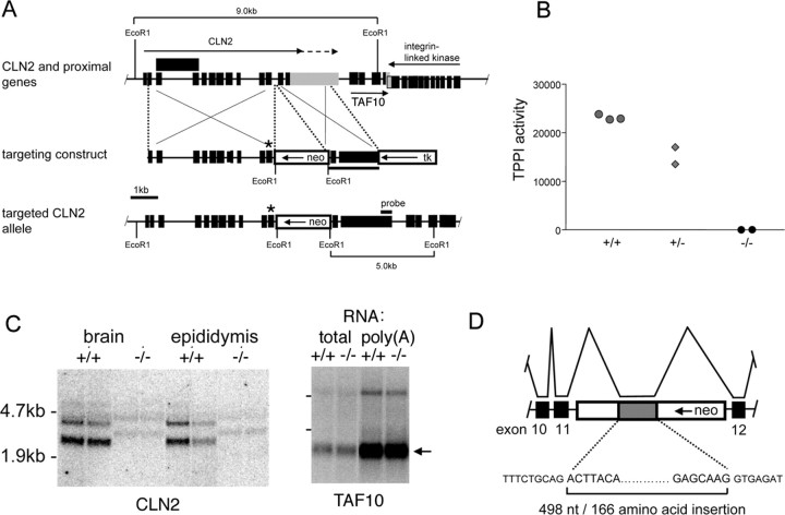
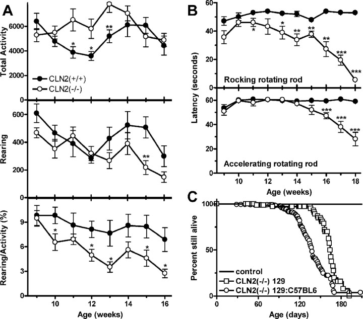
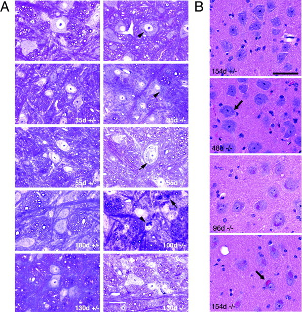
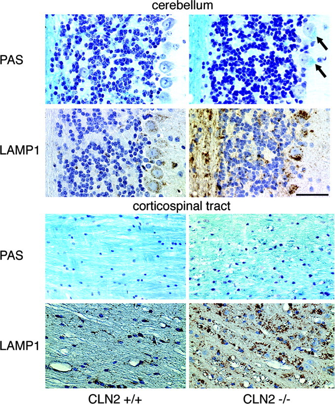
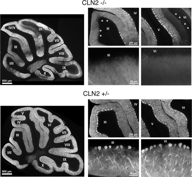
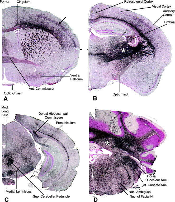
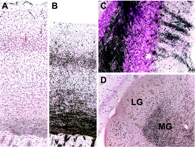
Mutations in the CLN2 gene, which encodes a lysosomal serine protease, tripeptidyl-peptidase I (TPP I), result in an autosomal recessive neurodegenerative disease of children, classical late-infantile neuronal ceroid lipofuscinosis (cLINCL). cLINCL is inevitably fatal, and there currently exists no cure or effective treatment. In this report, we provide the characterization of the first CLN2-targeted mouse model for cLINCL. CLN2-targeted mice were fertile and apparently healthy at birth despite an absence of detectable TPP I activity. At approximately 7 weeks of age, neurological deficiencies became evident with the onset of a tremor that became progressively more severe and was eventually accompanied by ataxia. Lifespan of the affected mice was greatly reduced (median survival, 138 d), and extensive neuronal pathology was observed including a prominent accumulation of cytoplasmic storage material within the lysosomal-endosomal compartment, a loss of cerebellar Purkinje cells, and widespread axonal degeneration. The CLN2-targeted mouse therefore recapitulates much of the pathology and clinical features of cLINCL and represents an animal model that should provide clues to the normal cellular function of TPP I and the pathogenic processes that underlie neuronal death in its absence. In addition, the CLN2-targeted mouse also represents a valuable model for the evaluation of different therapeutic strategies.


Figures

Figure 1.
Figure 1.
Targeted disruption of the mouse CLN2 gene. A, Structure of CLN2 and proximal genes, the targeting construct, and the targeted CLN2 locus (boxes: black shading, exons; gray shading, CLN2 3′-untranslated sequence; unshaded, thymidine kinase and neo selection markers). Murine CLN2 transcripts terminate at two different polyadenylylation sites (indicated by arrows above CLN2 gene), and in some cases, intron 3 is not spliced from the mature mRNA (the structure of the corresponding transcript is indicated by an extended exon box above the CLN2 gene). The targeted CLN2 gene is disrupted by the insertion of neo into intron 11 and by the incorporation of an Arg446His missense mutation into exon 11 immediately upstream of the neo insertion (indicated by *). The integrin linked kinase gene is set lower than TAF10 because they are overlapping. B, TPP I enzyme assays in wild-type, heterozygote, and homozygous CLN2-targeted mice; each point represents an individual mouse. C, Detection of CLN2 and TAF10 mRNAs in duplicate mutant and control mice by Northern blotting. Note the CLN2 transcripts of ∼500 nt greater length than the two wild-type mRNAs that are weakly detectable in the -/- mouse. Full-length TAF10 mRNA is ∼0.8 kb, and this size is consistent with the major transcript detected (arrow). D, Schematic of the aberrant splicing event that introduces 498 additional nucleotides derived from the neo selection marker to the CLN2 mRNA.
Figure 2.
Figure 2.
Survival and motor function phenotype in the CLN2 mutant. A, Open-field motor activity and rearing measurements on wild-type (filled symbols; n = 8-11) and CLN2(-/-) (open symbols; n = 8-11) animals. B, Motor coordination and balance measurements on wild-type (n = 10) and CLN2(-/-) (n = 9) animals using accelerating and rocking versions of the rotating rod test. C, Survival analysis of CLN2 mutants and controls in a mixed 129Sv/C57BL6 background and in an isogenic 129Sv background. For the mixed 129Sv/C57BL6 background (-/-) (n = 286), control mice were heterozygous (n = 226) and CLN2 (+/+) (n = 124) mice in both mixed-strain and isogenic backgrounds. In the mixed and isogenic backgrounds, 286 and 37 -/- mice were analyzed, respectively. For A and B, data are mean ± SEM. *p < 0.05; **p < 0.01; ***p < 0.001.
Figure 3.
Figure 3.
Unstained paraffin-processed sections of neocortex photographed under epifluorescence from +/- control (A) and -/- (B) at 154 d survival and demonstrating a significant increase in autofluorescent lysosomes within the mutant brain.
Figure 4.
Figure 4.
Progressive accumulation of cytoplasmic storage material in CLN2-targeted mouse brain. Ages of CLN2 mutants and age-matched littermate heterozygote controls are indicated on each panel. A, PAS-stained sections from the brainstem at the level of the hypoglossal nucleus. Brains were processed through JB-4 plastic embedding media. Positive inclusions in the cell cytoplasm are evident (punctate granules; arrowheads). Arrow in 100 d -/- panel indicates the presence of inclusions in neural fibers. Scale bar, 20 μm. B, H&E-stained sections of large neurons in the reticular nucleus of the medulla. Neuronal eosinophilic inclusions are barely visible at 48 d (arrow) but increase significantly in number and size with survival time. By 154 d survival, the inclusions appear to have aggregated into a single mass in some neurons (arrow). Neuronal eosinophilic inclusions were not present in tissue from littermate controls (+/+ or +/-) (non-neuronal staining derives from residual erythrocytes not removed during perfusion). Scale bar, 50 μm.
Figure 5.
Figure 5.
Matched sections from the cerebellum and corticospinal tract stained for LFB-CV and immunohistochemically for LAMP-1. In the cerebellum, there is a loss of Nissl substance from -/- cells, and storage pathology is visible as bright blue deposits within the cytoplasm of Purkinje cells (arrows). In both the corticospinal tract and cerebellum, lysosomal staining for LAMP-1 is markedly increased. In the latter, staining in increased in both Purkinje cells of -/- animals and in the underlying cerebellar white matter. Control mice (+/+) were 104 or 154 d old, and mutants (-/-) were 115 or 154 d old. Scale bar, 50 μm.
Figure 6.
Figure 6.
Electron photomicrographs of cells in sections of cortex from CLN2-targeted mice at different ages and a 100 d +/- control littermate. Lysosomal curvilinear bodies are indicated by asterisks. Tissues were processed in Spurr's media, and 0.05 μm sections analyzed by transmission electron microscopy. Scale bar, 500 nm.
Figure 7.
Figure 7.
Degenerative changes affecting cerebellar Purkinje cells. Cerebellar sections from a 77-d-old CLN2-targeted mouse (top set) and heterozygote controls (bottom set) were stained for calbindin and photographed at low magnification (large left panel of each set) showing a patchy loss of Purkinje cells and associated dendrites within the molecular layer. Lobes III and VI, which showed significant loss of Purkinje cells in the mutant (asterisks), were photographed at medium magnification (top right panels of each set), and the boundaries between the molecular and granular layers were photographed at high magnification (bottom right panels of each set).
Figure 8.
Figure 8.
Rostrocaudal series of sections through the brain of a male -/- animal at 132 d survival stained by the DeOlmos silver degeneration method. A, Rostrally, at the level of the decussation of the anterior commissure, impregnation of structures is anatomically discrete. The anterior limb of the anterior commissure is moderately stained. The cingulum and fiber bundles crossing the caudate-putamen are strongly stained, as is the fornix. The ventral pallidum, but not the adjacent pyriform cortex (asterisk), is moderately positive. In the cerebral neocortex, the somatosensory cortex is distinctly positive (arrow), but ventral allocortex is relatively negative with a fairly sharp boundary between them (arrowhead). The optic chiasm is unstained. B, At the level of the diencephalon, the hypothalamus is unstained, but the lateral thalamus is strongly positive (asterisk), as are fiber pathways connecting thalamus with cerebral cortex. The optic tract, in contrast, is virtually unstained. A discrete area of mossy fiber degeneration is present in the hippocampus (arrow). Within the cerebral cortex, the auditory cortex is positive, particularly in layer 4 and deeper layer 6, whereas the adjacent visual cortex is almost negative. Staining of the retrosplenial cortex is slight. C, At the midbrain level, several tracts are intensely stained including the dorsal hippocampal commissure, medial longitudinal fasciculus, medial lemniscus, and superior cerebellar peduncle. D, Section through cerebellum and medulla. Groups of stained Purkinje cells in parasagittal bands are visible in some cerebellar cortical lobules such as the simplex (arrows; see also Fig. 9C). Heavily stained Purkinje axons fill the cerebellar white matter and terminate on mostly unstained neurons in the cerebellar nuclei (asterisk). Additional strong staining, mainly of axon terminals, is seen in the lateral cuneate and dorsal cochlear nuclei. Cell bodies, but not their axons, are strongly stained in the motor nucleus of the facial nerve and nucleus ambiguus. Representative results are shown from silver staining of four CLN2 mutants and two age-matched controls.
Figure 9.
Figure 9.
Higher-magnification illustrations from mouse brain sections stained by the DeOlmos silver degeneration method. A, Somatosensory cortex in a +/+ control. Staining is limited to a light, nonspecific “dusting” overlying cell nuclei and intense reaction within poorly perfused blood vessels. B, The corresponding somatosensory regions of a CLN2 mutant. Fibers throughout the striatum, callosum, and neocortex are intensely impregnated. The neocortex is also noticeably thinner at this level compared with control. C, In the cerebellar cortex of a CLN2 mutant, a cluster of Purkinje cells is densely stained (cell bodies along with their dendritic trees extending through the molecular layer), as well as fibers in the underlying white matter. The cell body of the lowermost Purkinje cell is arrowed. D, As an example of anatomical selectivity in silver staining, fibers coursing into the medial geniculate (MG) are strongly positive in a CLN2 mutant, as are most other components in the auditory circuitry, whereas the adjacent lateral geniculate (LG) that receives visual projections is unstained (except for nonperfused blood vessels). Mice were male and 128-132 d of age.

References

    1. Bronson RT, Donahue LR, Johnson KR, Tanner A, Lane PW, Faust JR (1998) Neuronal ceroid lipofuscinosis (nclf), a new disorder of the mouse linked to chromosome 9. Am J Med Genet 77: 289-297. - PubMed
    1. Chen JW, Pan W, D'Souza MP, August JT (1985) Lysosome-associated membrane proteins: characterization of LAMP-1 of macrophage P388 and mouse embryo 3T3 cultured cells. Arch Biochem Biophys 239: 574-586. - PubMed
    1. DeOlmos JS, Ingram WR (1971) An improved cupric-silver method for impregnation of axonal and terminal degeneration. Brain Res 33: 523-529. - PubMed
    1. Elleder M, Franc J, Kraus J, Nevsimalova S, Sixtova K, Zeman J (1997) Neuronal ceroid lipofuscinosis in the Czech Republic: analysis of 57 cases. Report of the `Prague NCL group'. Eur J Paediatr Neurol 1: 109-114. - PubMed
    1. Gao H, Boustany RM, Espinola JA, Cotman SL, Srinidhi L, Antonellis KA, Gillis T, Qin X, Liu S, Donahue LR, Bronson RT, Faust JR, Stout D, Haines JL, Lerner TJ, MacDonald ME (2002) Mutations in a novel CLN6-encoded transmembrane protein cause variant neuronal ceroid lipofuscinosis in man and mouse. Am J Hum Genet 70: 324-335. - PMC - PubMed

Publication types

MeSH terms