

A Korean case of juvenile muscular atrophy of distal upper extremity (Hirayama disease) with dynamic cervical cord compression
- PMID: 15483361
- PMCID: PMC2816348
- DOI: 10.3346/jkms.2004.19.5.768
A Korean case of juvenile muscular atrophy of distal upper extremity (Hirayama disease) with dynamic cervical cord compression
Abstract
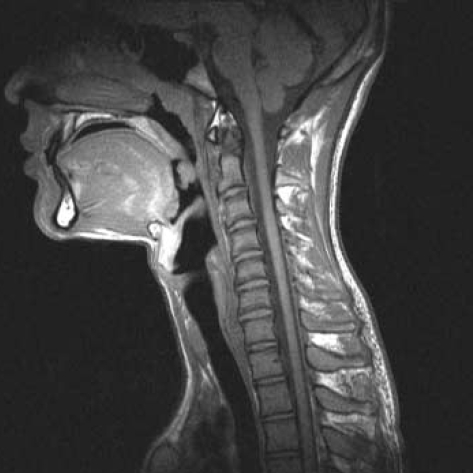
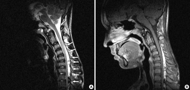
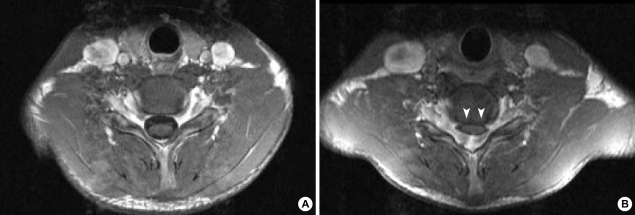
We present a Korean case of Hirayama disease with its typical neuroradiological findings of forward displacement of cervical dural sac and compression of the lower cervical cord during neck flexion. A 15-yr-old boy was presented with a one-year history of progressive weakness and atrophy affecting bilateral hands and forearms. The electrodiagnostic findings were compatible with the lesion of the anterior horn cells at the C7, C8, and T1 spinal segments. With neck flexion, cervical magnetic resonance imaging (MRI) showed the anterior shifting of the lower cervical dural sac resulting in the cord compression of those segments. Presumably, this disease might have been prevalent in Korea frequently under the diagnosis of "benign focal amyotrophy". In this regard, we discuss the clinical importance of cervical MRI with neck flexion and anticipate the increasing reports of the case substantiated by its characteristic radiological features.
Figures
References
-
- Hirayama K, Toyokura Y, Tsubaki T. Juvenile muscular atrophy of unilateral upper extremity; a new clinical entity. Psychiatr Neurol Jpn. 1959;61:2190–2197. - PubMed
-
- Pradhan S, Gupta RK. Magnetic resonance imaging in juvenile asymmetric segmental spinal muscular atrophy. J Neurol Sci. 1997;146:133–138. - PubMed
-
- Choi IS. Two cases of juvenile muscular atrophy confined to upper limb. J Korean Med Assoc. 1982;25:669–670.
-
- Choi IS. Benign focal amyotrophy. J Korean Med Assoc. 1987;30:1371–1374.
Publication types
MeSH terms
LinkOut - more resources
Full Text Sources
Medical
Miscellaneous
