. 2004 Nov 10;23(22):4423-33.
doi: 10.1038/sj.emboj.7600443. Epub 2004 Oct 14.

Latrophilin fragments behave as independent proteins that associate and signal on binding of LTX(N4C)

Affiliations

Latrophilin fragments behave as independent proteins that associate and signal on binding of LTX(N4C)

Kirill E Volynski et al. EMBO J. .

Abstract

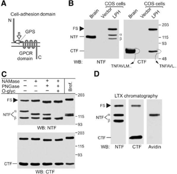
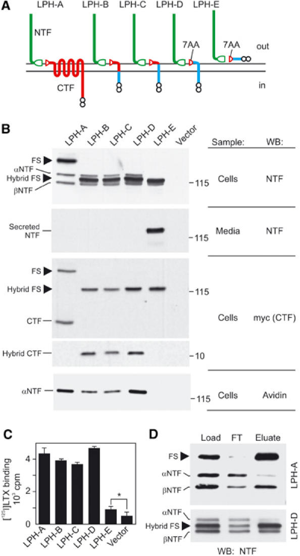
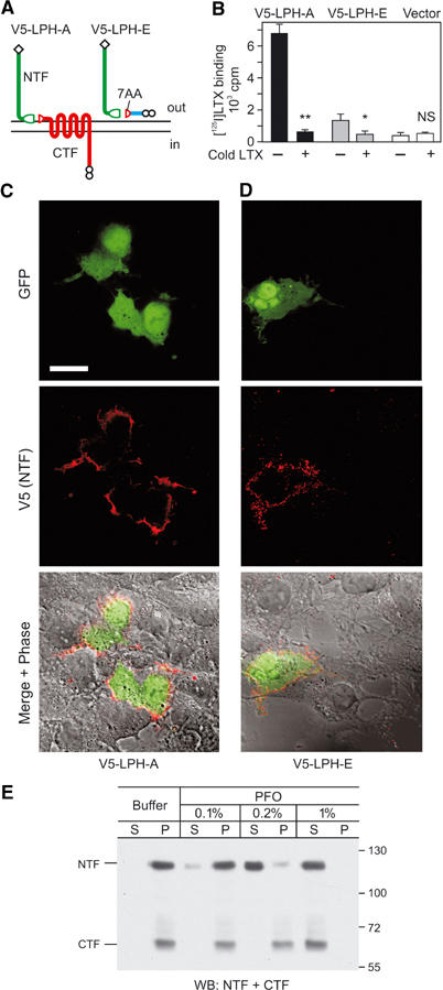
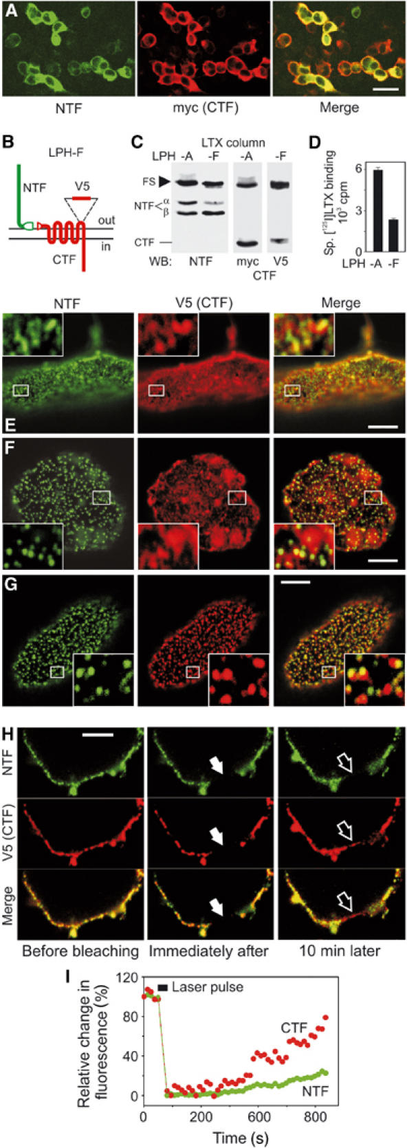
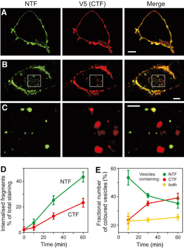
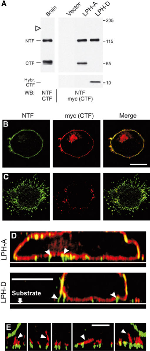
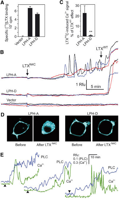
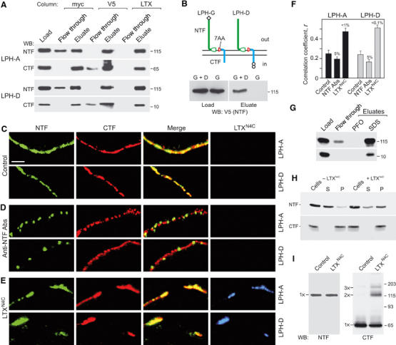
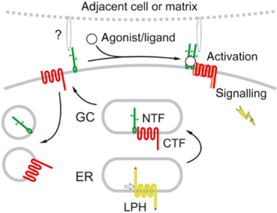
Heptahelical, or G-protein-coupled, receptors control many cellular functions and normally consist of one polypeptide chain. In contrast, heptahelical receptors that belong to the long N-terminus, group B (LNB) family are cleaved constitutively into two fragments. The N-terminal fragments (NTFs) resemble cell-adhesion proteins and the C-terminal fragments (CTFs) are typical G-protein-coupled receptors (GPCRs) with seven transmembrane regions. However, the functional roles of this cleavage and of any subsequent NTF-CTF interactions remain to be identified. Using latrophilin, a well-studied member of the LNB family, we now demonstrate that cleavage is critical for delivery of this receptor to the cell surface. On the plasma membrane, NTF and CTF behave as separate membrane proteins involved, respectively, in cell-surface reception and signalling. The two fragments can also internalise independently. However, separated NTF and CTF can re-associate on solubilisation. Agonist binding to NTF on the cell surface also induces re-association of fragments and provokes signal transduction via CTF. These findings define a novel principle of structural and functional organisation of the cleaved, two-subunit GPCRs.


Figures

Figure 1
Figure 1
Cellular processing of latrophilin. (A) A typical LNB GPCR. Constitutive cleavage (arrow) creates two fragments corresponding to the cell-adhesion domain and GPCR domain. (B) Analysis of latrophilin expression in COS7 cells. Solubilised receptors from rat brain and COS7 cells transfected with vector or LPH were enriched by α-latrotoxin chromatography and analysed by WB. N-terminal sequences of the native and recombinant CTFs extracted from the gel are shown. (C) Analysis of post-translational modification of latrophilin. Cells were either cultured in the presence of brefeldin A or solubilised and treated with glycosidases, as indicated. For abbreviations of enzymes, see Materials and methods. (D) Identification of surface-exposed species of latrophilin. Live cells were biotinylated, solubilised, enriched on α-latrotoxin column and analysed by WB. Only αNTF is labelled with biotin. Molecular masses are shown on the right (B, C). FS, full-size latrophilin.
Figure 2
Figure 2
Structural requirements for latrophilin cleavage and membrane anchoring. (A) Latrophilin constructs used herein. Green, NTF of latrophilin; red, remaining original CTF sequences; blue, foreign sequence; split ellipse, cleaved GPS domain; small circles, myc epitopes. (B) Analysis of expression, cleavage, secretion and surface exposure of the LPH constructs. Transfected COS7 cells were biotinylated, solubilised, enriched on α-latrotoxin columns and analysed by WB, as indicated, in parallel with conditioned medium. Molecular masses are shown on the right. (C) Binding of radiolabelled α-latrotoxin to cells expressing LPH constructs; *P<1%, t-test. (D) Immunoprecipitation of solubilised recombinant receptors with anti-myc mAb. FT, flow-through; FS, full-size latrophilin.
Figure 3
Figure 3
NTF can be anchored in the plasma membrane independently of CTF. (A) Modification of LPH-A and -E for improved immunodetection. Diamonds, N-terminal V5 epitopes. (B) Binding of [125I]-α-latrotoxin to COS7 cells transfected with V5-tagged constructs in the absence or presence of a 100-fold excess of cold toxin; **P<0.1%; *P<1%; NS, nonsignificant. (C, D) Immunostaining of V5-tagged constructs on the cell surface. COS7 cells co-transfected with GFP and V5-LPH-A (C) or V5-LPH-E (D) were stained with anti-V5 mAb. Detector gain for the red channel was increased in (D). Scale bar, 20 μm. (E) WB analysis of LPH-A-expressing neuroblastoma cells (see Figure 6 and respective text) treated with buffer or PFO at the indicated concentrations and centrifuged; S, supernatants; P, pellets. Note that 0.2% PFO solubilises only NTF but not CTF.
Figure 4
Figure 4
Latrophilin fragments behave as independent membrane proteins. (A) Indirect immunofluorescence of COS7 cells expressing LPH-A. (B) LPH-F with a surface-tagged CTF. (C) WB analysis of COS7 cells expressing LPH-A or -F. (D) Binding of radiolabelled α-latrotoxin to transfected cells. (E–G) Confocal images of apical membranes of nonpermeabilised cells stained for latrophilin fragments. Cells expressing LPH-F were: fixed and stained for NTF and CTF (E); or crosslinked with the primary and fluorescent secondary Abs against NTF, then stained for CTF (F); or crosslinked with primary and fluorescent secondary Abs against both fragments (G). Insets, enlarged boxed areas. NTF and CTF are independently re-arranged by crosslinking with respective Abs. (H) FRAP analysis of the lateral mobility of NTF and CTF. Cells expressing LPH-F were decorated with fluorescent primary Abs and imaged before and after photobleaching. Solid arrows, bleached area; open arrows, same area after recovery. (I) Quantification of NTF and CTF diffusion into the bleached area. Scale bars, 50 (A) and 5 (E–H) μm.
Figure 5
Figure 5
NTF and CTF can be separately internalised. (A, B) COS7 cells expressing LPH-F were decorated with both primary Abs and fixed before (A) or 30 min after (B) induction of endocytosis, then permeabilised and stained with secondary Abs. (C) Higher-magnification images of boxed areas in (B). NTF and CTF are found mostly in different vesicles. Scale bars, 5 (A, B) and 2 (C) μm. (D) Time courses of NTF and CTF endocytosis. LPH-F cells were stained as in (B), and the intensity of internalised fluorescence was measured relative to total fluorescence for each Ab. (E) Number of endocytosed vesicles containing NTF, CTF or both fragments. The data in (D, E) are the means±s.e.m., n=5–8.
Figure 6
Figure 6
NTF associates with cell-adhesion structures. (A) WB analysis of LPH-A and -D expression in stably transfected NB2a cells, compared to brain latrophilin. Open arrowhead, expected position of full-size LPH-A. (B, C) Distribution of NTF and CTF in NB2a cells expressing LPH-A. Horizontal confocal sections were imaged near the cell's middle (B) or near the substrate (C). (D) Vertical cross-sections reconstructed from a set of overlapping horizontal confocal sections, as in (B), for cells expressing LPH-A or -D. Arrowheads, cell-adhesion structures. (E) Association of actin fibres (red; arrowheads) with NTF-containing membrane protrusions (green) in vertical cross sections reconstructed as in (D) for several individual loci of two cells. Scale bars, 10 (B–D) and 4 (E) μm.
Figure 7
Figure 7
Latrotoxin induces CTF-mediated signalling. (A) Binding of [125I]-LTXN4C to NB2a cells expressing LPH-A or -D. (B) Time course of intracellular Ca2+ fluorescence in individual NB2a cells transfected with LPH-A, -D or vector and stimulated by 2.5 nM LTXN4C and native α-latrotoxin (LTXWT) (arrows). Rfu, relative fluorescence units. (C) Normalised maximal effect of LTXN4C; data are the means±s.e.m.; **P<0.1% (n=23–27 cells for each condition). (D) PLC activity visualised by translocation of CFP-PH into the cytosol after a 30-min application of 2.5 nM LTXN4C to cells expressing LPH-A or -D. (E) Time course of intracellular Ca2+ fluorescence and PLC activity in three individual LPH-A cells stimulated with LTXN4C at indicated times (arrowheads).
Figure 8
Figure 8
Molecular interaction between latrophilin fragments and role of LTXN4C. (A) Differential co-purification on different affinity columns of LPH fragments solubilised from NB2a cells transfected with LPH-A or LPH-D. (B) NTF from LPH-G (top) is pulled down with CTF of LPH-D by anti-myc column (bottom) upon solubilisation of NB2a cells co-expressing both constructs. (C–E) High-magnification confocal images of plasma membranes near the equator of NB2a cells expressing LPH-A or -D. Cells were fixed and stained without any treatments (C) or crosslinked with Abs against NTF (D), or treated with 2.5 nM fluorescent LTXN4C (E) before fixation and immunostaining. Scale bar, 2 μm. LTXN4C stimulates association of latrophilin fragments, while Abs crosslink NTF only. (F) Quantification of NTF and CTF co-localisation on the cell membranes. The data are the means±s.e.m. (n=25–30 cells for each condition). (G) 0.6% PFO does not break NTF–CTF complexes of LPH-D immobilised on anti-myc column, as in (A). (H) Pre-treatment of LPH-D-expressing cells with 5 nM LTXN4C inhibits solubilisation of NTF with 0.2% PFO. S, supernatant; P, pellet. (I) WB analysis of CTF oligomerisation in LTXN4C-stimulated NB2a cells expressing LPH-A.
Figure 9
Figure 9
Proposed scheme of LPH processing and activation (see text).

References

    1. Abe J, Fukuzawa T, Hirose S (2002) Cleavage of Ig-Hepta at a ‘SEA' module and at a conserved G protein-coupled receptor proteolytic site. J Biol Chem 277: 23391–23398 - PubMed
    1. Ashton AC, Volynski KE, Lelianova VG, Orlova EV, Van Renterghem C, Canepari M, Seagar M, Ushkaryov YA (2001) Latrotoxin, acting via two Ca2+-dependent pathways, triggers exocytosis of two pools of synaptic vesicles. J Biol Chem 276: 44695–44703 - PubMed
    1. Bouvier M (2001) Oligomerization of G-protein-coupled transmitter receptors. Nat Rev Neurosci 2: 274–286 - PubMed
    1. Capogna M, Volynski KE, Emptage NJ, Ushkaryov YA (2003) The α-latrotoxin mutant LTXN4C enhances spontaneous and evoked transmitter release in CA3 pyramidal neurons. J Neurosci 23: 4044–4053 - PMC - PubMed
    1. Chang GW, Stacey M, Kwakkenbos MJ, Hamann J, Gordon S, Lin HH (2003) Proteolytic cleavage of the EMR2 receptor requires both the extracellular stalk and the GPS motif. FEBS Lett 547: 145–150 - PubMed

Publication types

MeSH terms

LinkOut - more resources