Essential role for mitochondrial thioredoxin reductase in hematopoiesis, heart development, and heart function
- PMID: 15485910
- PMCID: PMC522221
- DOI: 10.1128/MCB.24.21.9414-9423.2004
Essential role for mitochondrial thioredoxin reductase in hematopoiesis, heart development, and heart function
Abstract
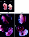
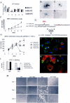
Oxygen radicals regulate many physiological processes, such as signaling, proliferation, and apoptosis, and thus play a pivotal role in pathophysiology and disease development. There are at least two thioredoxin reductase/thioredoxin/peroxiredoxin systems participating in the cellular defense against oxygen radicals. At present, relatively little is known about the contribution of individual enzymes to the redox metabolism in different cell types. To begin to address this question, we generated and characterized mice lacking functional mitochondrial thioredoxin reductase (TrxR2). Ubiquitous Cre-mediated inactivation of TrxR2 is associated with embryonic death at embryonic day 13. TrxR2(TrxR2(-/-)minus;/TrxR2(-/-)minus;) embryos are smaller and severely anemic and show increased apoptosis in the liver. The size of hematopoietic colonies cultured ex vivo is dramatically reduced. TrxR2-deficient embryonic fibroblasts are highly sensitive to endogenous oxygen radicals when glutathione synthesis is inhibited. Besides the defect in hematopoiesis, the ventricular heart wall of TrxR2(TrxR2(-/-)minus;/TrxR2(-/-)minus;) embryos is thinned and proliferation of cardiomyocytes is decreased. Cardiac tissue-restricted ablation of TrxR2 results in fatal dilated cardiomyopathy, a condition reminiscent of that in Keshan disease and Friedreich's ataxia. We conclude that TrxR2 plays a pivotal role in both hematopoiesis and heart function.
Figures


Similar articles
-
Cytoplasmic thioredoxin reductase is essential for embryogenesis but dispensable for cardiac development.Mol Cell Biol. 2005 Mar;25(5):1980-8. doi: 10.1128/MCB.25.5.1980-1988.2005. Mol Cell Biol. 2005. PMID: 15713651 Free PMC article.
-
Induction of apoptosis by the overexpression of an alternative splicing variant of mitochondrial thioredoxin reductase.Free Radic Biol Med. 2005 Dec 15;39(12):1666-75. doi: 10.1016/j.freeradbiomed.2005.08.008. Epub 2005 Sep 1. Free Radic Biol Med. 2005. PMID: 16298692
-
Oxidation of thioredoxin reductase in HeLa cells stimulated with tumor necrosis factor-alpha.FEBS Lett. 2004 Jun 4;567(2-3):189-96. doi: 10.1016/j.febslet.2004.04.055. FEBS Lett. 2004. PMID: 15178321
-
[Reactive oxygen species in regulation of cell migration. The role of thioredoxin reductase].Postepy Biochem. 2009;55(2):145-52. Postepy Biochem. 2009. PMID: 19824470 Review. Polish.
-
Significance of the mitochondrial thioredoxin reductase in cancer cells: An update on role, targets and inhibitors.Free Radic Biol Med. 2018 Nov 1;127:62-79. doi: 10.1016/j.freeradbiomed.2018.03.043. Epub 2018 Mar 27. Free Radic Biol Med. 2018. PMID: 29596885 Review.
Cited by
-
Mitochondria: Much ado about nothing? How dangerous is reactive oxygen species production?Int J Biochem Cell Biol. 2015 Jun;63:16-20. doi: 10.1016/j.biocel.2015.01.021. Epub 2015 Feb 7. Int J Biochem Cell Biol. 2015. PMID: 25666559 Free PMC article. Review.
-
The role of selenium metabolism and selenoproteins in cartilage homeostasis and arthropathies.Exp Mol Med. 2020 Aug;52(8):1198-1208. doi: 10.1038/s12276-020-0408-y. Epub 2020 Aug 13. Exp Mol Med. 2020. PMID: 32788658 Free PMC article. Review.
-
The Function of Selenium in Central Nervous System: Lessons from MsrB1 Knockout Mouse Models.Molecules. 2021 Mar 4;26(5):1372. doi: 10.3390/molecules26051372. Molecules. 2021. PMID: 33806413 Free PMC article. Review.
-
Epitranscriptomic systems regulate the translation of reactive oxygen species detoxifying and disease linked selenoproteins.Free Radic Biol Med. 2019 Nov 1;143:573-593. doi: 10.1016/j.freeradbiomed.2019.08.030. Epub 2019 Aug 30. Free Radic Biol Med. 2019. PMID: 31476365 Free PMC article. Review.
-
Redox signaling in cardiac myocytes.Free Radic Biol Med. 2011 Apr 1;50(7):777-93. doi: 10.1016/j.freeradbiomed.2011.01.003. Epub 2011 Jan 12. Free Radic Biol Med. 2011. PMID: 21236334 Free PMC article. Review.
References
-
- Arner, E. S., and A. Holmgren. 2000. Physiological functions of thioredoxin and thioredoxin reductase. Eur. J. Biochem. 267:6102-6109. - PubMed
-
- Chen, C. S., and K. R. Gee. 2000. Redox-dependent trafficking of 2,3,4,5,6-pentafluorodihydrotetramethylrosamine, a novel fluorogenic indicator of cellular oxidative activity. Free Radic. Biol. Med. 28:1266-1278. - PubMed
-
- Chen, J., S. W. Kubalak, and K. R. Chien. 1998. Ventricular muscle-restricted targeting of the RXRα gene reveals a non-cell-autonomous requirement in cardiac chamber morphogenesis. Development 125:1943-1949. - PubMed
-
- Conrad, M., M. Brielmeier, W. Wurst, and G. W. Bornkamm. 2003. Optimized vector for conditional gene targeting in mouse embryonic stem cells. BioTechniques 34:1136-1140. - PubMed
Publication types
MeSH terms
Substances
LinkOut - more resources
Full Text Sources
Molecular Biology Databases
