. 2004 Nov;24(21):9437-55.
doi: 10.1128/MCB.24.21.9437-9455.2004.

Interactions of eukaryotic translation initiation factor 3 (eIF3) subunit NIP1/c with eIF1 and eIF5 promote preinitiation complex assembly and regulate start codon selection

Affiliations

Interactions of eukaryotic translation initiation factor 3 (eIF3) subunit NIP1/c with eIF1 and eIF5 promote preinitiation complex assembly and regulate start codon selection

Leos Valásek et al. Mol Cell Biol. 2004 Nov.

Abstract

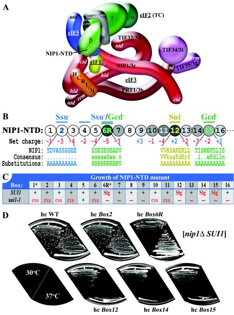
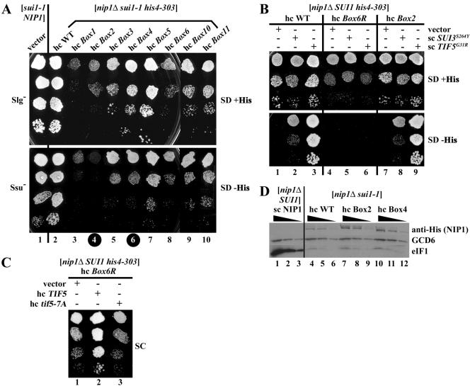
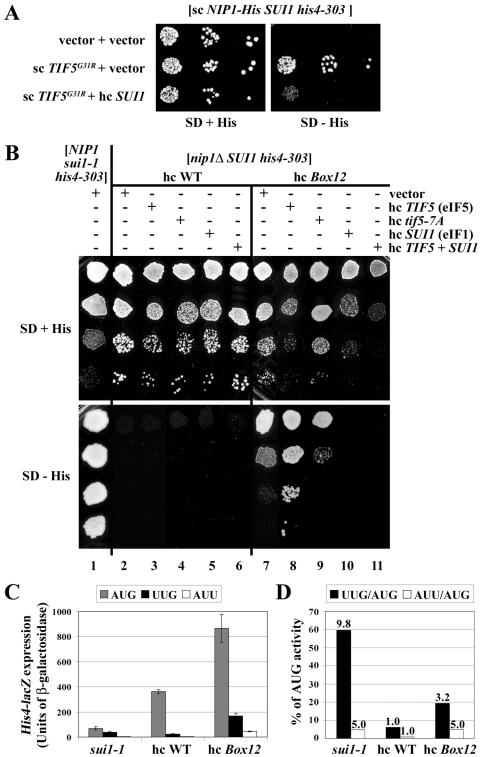
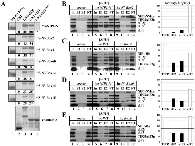
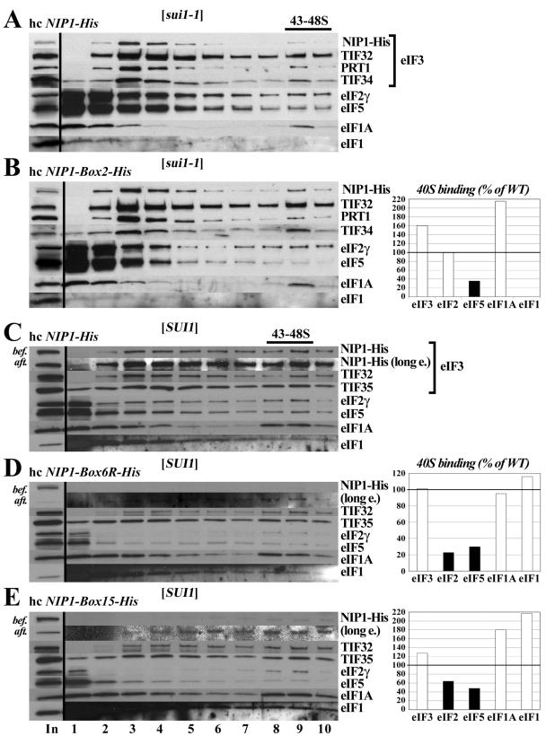
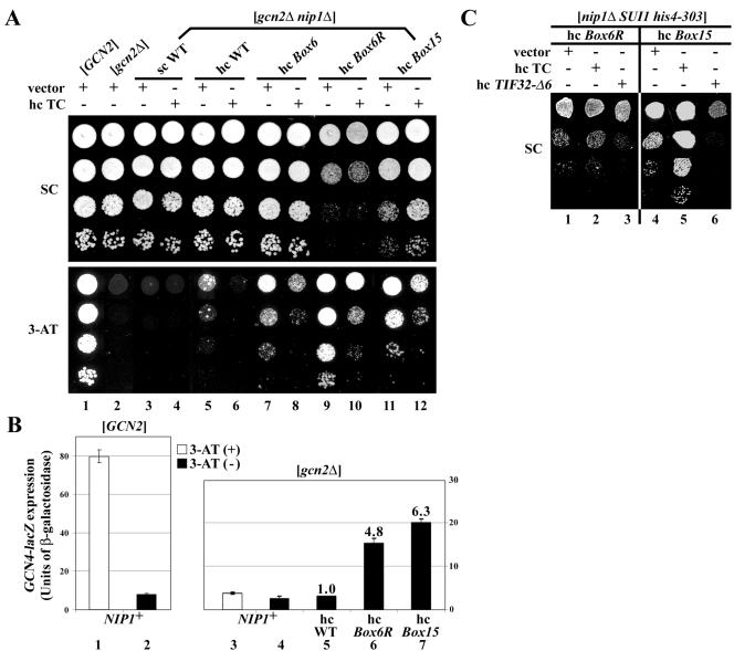
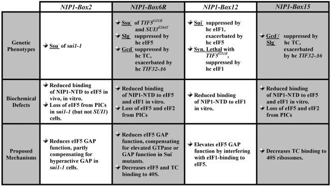
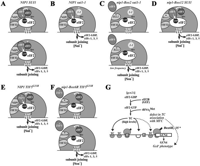
The N-terminal domain (NTD) of NIP1/eIF3c interacts directly with eIF1 and eIF5 and indirectly through eIF5 with the eIF2-GTP-Met-tRNA(i)(Met) ternary complex (TC) to form the multifactor complex (MFC). We investigated the physiological importance of these interactions by mutating 16 segments spanning the NIP1-NTD. Mutations in multiple segments reduced the binding of eIF1 or eIF5 to the NIP1-NTD. Mutating a C-terminal segment of the NIP1-NTD increased utilization of UUG start codons (Sui(-) phenotype) and was lethal in cells expressing eIF5-G31R that is hyperactive in stimulating GTP hydrolysis by the TC at AUG codons. Both effects of this NIP1 mutation were suppressed by eIF1 overexpression, as was the Sui(-) phenotype conferred by eIF5-G31R. Mutations in two N-terminal segments of the NIP1-NTD suppressed the Sui(-) phenotypes produced by the eIF1-D83G and eIF5-G31R mutations. From these and other findings, we propose that the NIP1-NTD coordinates an interaction between eIF1 and eIF5 that inhibits GTP hydrolysis at non-AUG codons. Two NIP1-NTD mutations were found to derepress GCN4 translation in a manner suppressed by overexpressing the TC, indicating that MFC formation stimulates TC recruitment to 40S ribosomes. Thus, the NIP1-NTD is required for efficient assembly of preinitiation complexes and also regulates the selection of AUG start codons in vivo.


Figures

FIG. 1.
FIG. 1.
Phenotypic analysis of the NIP1-NTD mutants. (A) Three-dimensional model of the MFC based on a comprehensive analysis of subunit interactions (29). The labeled protein subunits are shown roughly in proportion to their molecular weights. The degree of overlap between two different subunits depicts the extent of their interacting surfaces. The boundaries of the N terminus of NIP1 (NIP1-NTD) subjected to mutagenesis are indicated by dotted white lines. ntd, N-terminal domain; ctd, C-terminal domain; hld, HCR1-like domain; rrm, RNA recognition motif; TC, ternary complex. (B) The sequence of the first 160 amino acid residues of NIP1 is shown as numbered circles (boxes 1 to 16), each of them composed of 10 residues that were substituted by a stretch of 10 alanines. Different shades of gray indicate the degree of identities between the NIP1-NTD and the N termini of its Caenorhabditis elegans, Human sapiens, Arabidopsis thaliana, and Schizosaccharomyces pombe homologues that were aligned by using the GCG Sequence Analysis Program (version 8; Genetics Computer Group, Inc., Madison, Wis.) (white, <20%; light gray, 20 to 40%; medium gray, 40 to 60%; dark gray, 60 to 80%). Color-coded bars above the circles indicate the phenotypes associated with amino acid substitutions in the corresponding boxes: Ssu (suppressor of Sui), Gcd (general control derepressed), and Sui (suppressor of initiation codon). Blown-up segments in blue, green, and yellow indicate the amino acid sequences, a consensus sequence derived from sequence alignments, and the substitutions made in the corresponding boxes of the NIP1-NTD. (C) Summary of the growth phenotypes of the NIP1 mutants in a SUI1 strain (second row) and their genetic interactions with sui1-1 (third row). The indicated plasmid-borne hc NIP1-Box alleles (row 1) were introduced into strains HLV04 (nip1Δ SUI1) and HLV05 (nip1Δ sui1-1), both carrying WT NIP1 on a single-copy (sc) URA3 plasmid that was subsequently evicted by growth on 5-FOA medium. Synthetic lethality with sui1-1 was identified by the failure to grow on 5-FOA plates. Growth of the viable strains was analyzed by determining the sizes of colonies formed from single cells streaked on yeast extract-peptone-dextrose (YPD) plates. In the second row, “+” and Slg designate WT and slow-growth phenotypes, respectively, in the SUI1 strain. In the third row, “+”, “exa”, and “−” indicate no effect, exacerbation of the Slg- phenotype, and synthetic lethality in the sui1-1 strain, respectively. The asterisks in row 1 designate mutants that are lethal when expressed from a single-copy plasmid. (D) Growth phenotypes of selected NIP1 mutants. The strains derived from HLV04 (nip1Δ SUI1 his4-303) containing the indicated NIP1 alleles on high-copy-number (hc) plasmids were streaked for single colonies on yeast extract-peptone-dextrose medium at 30°C (left-hand sectors) and 37°C (right-hand sectors).
FIG. 2.
FIG. 2.
The Box2, Box4, and Box6R alleles of NIP1 exhibit Ssu (suppressor of Sui) phenotypes. (A) Suppression of the Sui phenotype of sui1-1 by the hc Box2 and hc Box4 alleles. Derivatives of HLV05 (nip1Δ sui1-1 his4-303) containing hc WT NIP1-His on plasmid YEpNIP1-His (lane 2) or the indicated hc NIP1 mutant alleles on the appropriate derivatives of YEpNIP1-His plasmids (lanes 3 to 10), and the parental strain TD301-8D (NIP1 sui1-1 his4-303) transformed with empty vector (lane 1), were spotted in four serial dilutions on SD medium containing histidine (upper panel) or lacking histidine (lower panel) and incubated at 30°C for 7 days. Black circles on the column numbers highlight the mutants displaying Ssu phenotypes. (B) NIP1-Box6R suppresses the dominant Sui phenotypes of SUI3-S264Y and TIF5-G31R. Derivatives of HLV04 (nip1Δ SUI1 his4-303) containing hc NIP1-His on YEpNIP1-His (lanes 1 to 3), hc NIP1-Box6R-His on YEpNIP1-Box6R-His (lanes 4 to 6), and hc NIP1-Box2-His on YEpNIP1-Box2-His (lanes 7 to 9) were transformed with empty vector YCplac33 (lanes 1, 4, and 7), lc plasmid pRSSUI3-S264Y-U harboring SUI3-S264Y (lanes 2, 5, and 8), and sc plasmid YCpTIF5-G31R-U harboring TIF5-G31R (lanes 3, 6, and 9). The resulting transformants were spotted in three serial dilutions on SD plates supplemented with histidine (upper panel) or lacking histidine (lower panel) and incubated at 30°C for 3 days. (C) Overexpression of eIF5 but not eIF5-7A suppresses the Slg phenotype of the hc NIP1-Box6R mutant. The derivative of HLV04 containing hc NIP1-Box6R-His was transformed with empty vector (lane 1), hc plasmid YEpTIF5-U harboring TIF5 (lane 2), or hc plasmid YEpTIF5-7A-U harboring tif5-7A (lane 3) and the resulting transformants were spotted in four serial dilutions on SC medium (containing histidine) and incubated at 30°C for 3 days. (D) The D83G substitution in eIF1 (sui1-1) reduces the steady-state level of eIF1. The derivative of HLV04 containing WT untagged NIP1 on sc plasmid pNIP1+ (lanes 1 to 3) and derivatives of HLV05 containing NIP1-His (lanes 4 to 6), NIP1-Box2-His (lanes 7 to 9), or NIP1-Box4-His (lanes 10 to 12) on hc plasmids were grown in YPD medium, and WCEs were subjected to Western analysis with antibodies against the His8 epitope to detect the NIP1-His proteins, or against GCD6 or eIF1, as indicated to the right of the panels. Three different dilutions of each WCE were loaded in consecutive lanes as indicated by the black triangles.
FIG. 3.
FIG. 3.
The NIP1-Box12 mutation produces a Sui phenotype that is suppressed by overexpression of eIF1 and exacerbated by overexpression of eIF5. (A) Overexpression of eIF1 suppresses the dominant Sui phenotype conferred by TIF5-G31R. The derivative of HLV04 (nip1Δ SUI1 his4-303) containing sc plasmid YCpNIP1-His-L harboring WT NIP1-His was transformed with the empty vectors YCplac22 and YEplac195 (row 1), with sc plasmid YCpTIF5-G31R-W harboring TIF5-G31R and YEplac195 (row 2), or with YCpTIF5-G31R-W and hc SUI1 plasmid YEpSUI1-U (row 3), and the resulting transformants were spotted in three serial dilutions on the SD plates supplemented with histidine (left-hand panel) or lacking histidine (right-hand panel) and incubated at 30°C for 5 days. (B) The HLV04-derivatives containing hc NIP1-His on YEpNIP1-His (lanes 2 to 6) or hc NIP1-Box12-His on YEpNIP1-Box12-His (lanes 7 to 11) were transformed with empty vector YEplac195 (lanes 2 and 7), hc TIF5 plasmid YEpTIF5-U (lanes 3 and 8), hc tif5-7A plasmid YEpTIF5-7A-U (lanes 4 and 9), hc SUI1 plasmid YEpSUI1-U (lanes 5 and 10), or hc plasmid YEpTIF5+SUI1 harboring TIF5 and SUI1 (lanes 6 and 11), and the resulting transformants and the parental strain TD301-8D (NIP1 sui1-1 his4-303) transformed with empty vector (lane 1) were spotted in four serial dilutions on SD medium containing histidine (upper panel) or lacking histidine (lower panel) and incubated at 30°C for 2 days (upper panel) or 7 days (lower panel). (C and D) HLV04 derivatives containing hc NIP1-His or hc NIP1-Box12-His and TD301-8D (NIP1 sui1-1 his4-303) were transformed with p367, p391, and p2042 containing, respectively, HIS4-lacZ reporters harboring ATG, TTG, and ATT start codons. The resulting transformants were grown in SC medium lacking uracil and tryptophan (SC−Ura−Trp) and WCE extracts were prepared and assayed for β-galactosidase activity as described previously (19). Panel C shows the averages and standard deviations from at least six independent measurements with three independent transformants. β-Galactosidase activity was expressed in nanomoles of o-nitrophenyl-β-d-galactopyranoside hydrolyzed per minute per milligram of protein. In panel D, the black bars in the histogram show the mean ratios of expression from the UUG to the AUG reporter, and white bars show the mean ratios of expression from the AUU to the AUG reporter. The fold increases in these two ratios measured in the sui1-1 and hc NIP1-Box12-His mutants versus WT are indicated above the corresponding bars.
FIG. 4.
FIG. 4.
Mutations in the NIP1-NTD impair its binding to eIF1 and eIF5. (A) In vitro binding assays. Full-length eIF5 (lane 3), eIF1 (lane 4), or eIF1-D83G (lane 5) fused to GST, as well as GST alone (lane 2), were expressed in E. coli, immobilized on glutathione-Sepharose beads and incubated with the indicated WT (top panel) or mutant 35S-labeled NIP1-NTD polypeptides synthesized in rabbit reticulocyte lysates. The beads were washed with phosphate-buffered saline, and the bound proteins were eluted, separated by sodium dodecyl sulfate-polyacrylamide gel electrophoresis, stained with Gelcode Blue Stain Reagent (Pierce) (bottom panel; Coomassie), and subjected to autoradiography (upper panels). Lane 1 shows 20% of the input amounts of in vitro-translated proteins added to each reaction (Input [20%]). The amount of each 35S-labeled NIP1 polypeptide bound to each GST-fusion protein was quantified and is expressed below the corresponding panel as a percentage of the binding observed for WT 35S-labeled NIP1. The mobilities of the 35S-labeled proteins in the input lanes are increased artifactually by the large amount of β-globin present in the reticulocyte lysate. (B) The NIP1-Box2 mutation diminishes binding of eIF5 and eIF2 to the NIP1-NTD in vivo. WCEs were prepared from transformants of strain HLV04 bearing empty vector (lanes 1 to 4), hc plasmid YEpNIP1-N′-His-X (lanes 5 to 8), or hc plasmid YEpNIP1-N′-Box2-His (lanes 9 to 12), with the latter two plasmids encoding WT or Box2 versions of the N-terminal 205 residues of NIP1. WCEs were incubated with Ni2+-nitrilotriacetic acid-silica resin, and the bound proteins were eluted and subjected to Western blot analysis with antibodies to the His8 epitope (to detect the NIP1-NTD polypeptides) or with antibodies to the other factors listed to the right of the blots. Lanes 1, 5, and 9 contained 3% of the input WCEs (In); lanes 2, 6, and 10 contained 15% of the first fractions eluted from the resin (E1); lanes 3, 7, and 11 contained 30% of the same fractions (E2); and lanes 4, 8, and 12 contained 3% of the flowthrough fractions (FT). The Western signals for eIF2, eIF1, and eIF5 in the E1 and E2 fractions for the Box2 mutant (lanes 10 to 11) were quantified, combined, normalized for the amounts of the NIP1-NTD-Box2 fragment in these fractions, and plotted in the histogram on the right as percentages of the corresponding values calculated for the WT NIP1-NTD (fractions 6 to 7). (C) The NIP1-Box2 mutation reduces association of eIF5 and eIF2 with the MFC in vivo. Same as in panel B except that WCEs were prepared from transformants of strain HLV04 bearing empty vector (lanes 1 to 4), hc plasmid YEpNIP1-His (lanes 5 to 8), or YEpNIP1-Box2-His (lanes 9 to 12), with the latter two plasmids encoding WT or Box2 versions of full-length NIP1-His. (D) The NIP1-Box6 mutation diminishes binding of eIF5 and eIF2 to the NIP1-NTD in vivo. Same as in panel B except that WCEs were prepared from transformants bearing empty vector (lanes 1 to 4), hc plasmid YEpNIP1-N′-His-X (lanes 5 to 8), or hc plasmid YEpNIP1-N′-Box6-His (lanes 9 to 12), with the latter two plasmids encoding WT or Box6 versions of the NIP1-NTD. (E) The NIP1-Box6 mutation reduces association of eIF5 and eIF2 with the MFC in vivo. Panel E is the same as panel B except that WCEs were prepared from transformants of HLV04 bearing empty vector (lanes 1 to 4), YEpNIP1-His (lanes 5 to 8), or YEpNIP1-Box6-His (lanes 9 to 12), with the latter two plasmids encoding WT or Box6 versions of full-length NIP1-His.
FIG. 5.
FIG. 5.
NIP1-NTD mutations impair the assembly of preinitiation complexes in vivo. (A and B) eIF5 binds poorly to 40S subunits in sui1-1 cells expressing NIP1-Box2-His. Derivatives of HLV05 (nip1Δ sui1-1 his4-303) containing hc NIP1-His (A) or hc NIP1-Box2-His (B) were grown in YPD medium and cross-linked with formaldehyde. WCEs were prepared and separated by velocity sedimentation on a sucrose gradient. The gradients were collected and scanned at 254 nm to visualize the ribosomal species. Proteins were subjected to Western analysis with antibodies to the proteins listed to the right of the blots or with antibodies to the His8 epitope to detect the NIP1-His proteins. The first lane contains a sample of the starting WCEs resolved on the gradients (In). The Western signals in fractions 9 and 10 containing the 43S-48S complexes were quantified, and the amounts of each factor from the NIP1-Box2-His extract are plotted in the histogram on the right of panel B as percentages of the corresponding amounts measured for the WT extract analyzed in panel A. (C to E) The Box6R and Box15 mutations reduce the amounts of eIF5 and eIF2 associated with 40S ribosomes in vivo. Same as panels A and B except that derivatives of HLV04 (nip1Δ SUI1 his4-303) expressing WT NIP1-His (C), NIP1-Box6R-His (D), or NIP1-Box15-His (E), respectively, were analyzed after growth in SD medium. Short and long (long e.) exposures are shown for the Western analysis of NIP1-His proteins in the gradient factions. In addition, samples of the input WCEs were subjected to Western analysis of the NIP1-His proteins immediately before (bef.) or after (aft.) incubation for 7 h on ice (lane 1, In).
FIG. 6.
FIG. 6.
Genetic evidence that the Box6R and Box15 NIP1-NTD mutations reduce the rate of TC recruitment to 40S ribosomes and thereby impair GCN4 translational control. (A) Derivatives of strain HKN06 (gcn2Δ nip1Δ) containing sc NIP1-His (lanes 3 and 4), hc NIP1-His (lanes 5 and 6), and the indicated hc NIP1-Box-His mutants (lanes 7 to 12) were transformed with empty vector YEplac195 (lanes with odd numbers beginning with lane 3) or hc plasmid p1780-IMT encoding all three subunits of eIF2 and tRNAiMet (hc TC) (lanes with even numbers beginning with lane 4). The resulting transformants and isogenic strains H2880 (GCN2) (lane 1) and H2881 (gcn2Δ) (lane2) were spotted in four serial dilutions on SC (upper panel) or SC containing 30 mM 3-AT (lower panel) and then incubated at 30°C for 5 days. (B) The strains described in panel A containing hc WT NIP1-His (lane 5), hc NIP1-Box6R-His (lane 6), or hc NIP1-Box15-His (lane 7), as well as GCN2 NIP1strain H2880 (lanes 1 and 2) and gcn2Δ NIP1 strain H2881 (lanes 3 and 4), were transformed with p180 containing the GCN4-lacZ fusion with all four uORFs present. The transformants were grown in SC−Ura in the presence of 10 mM 3-AT (white bars, lanes 1 and 3) or without 3-AT (black bars; lanes 2 and 4 to 7), and the β-galactosidase activities were measured in the WCEs. The histogram shows the mean values and standard deviations obtained from at least six independent measurements with three independent transformants. The fold increases observed in the hc NIP1-His mutants versus hc WT NIP1-His are given above the relevant bars. (C) The HLV04 derivatives (nip1Δ SUI1 his4-303) containing the indicated NIP1-Box-His mutants were transformed with empty vector (lanes 1 and 4), p1780 (lanes 2 and 5), or hc plasmid YEp-TIF32-Δ6-His-U containing TIF32-Δ6-His (lanes 3 and 6), and the resulting transformants were spotted in three serial dilutions on SC plates and incubated at 30°C for 3 days.
FIG. 7.
FIG. 7.
Summary of phenotypes, biochemical defects, and proposed mechanisms for the NIP1 mutants analyzed in the present study. Ssu (suppressor of Sui), Slg (slow-growth), Gcd (general control derepressed), Sui (suppressor of initiation codon), Syn. Lethal (synthetic lethal), PIC (preinitiation complex), GAP (GTP activating protein) are indicated. See Results and Discussion for further details.
FIG. 8.
FIG. 8.
Hypothetical models depicting the proposed functions of eIF1, eIF3, eIF5, and TC in AUG selection and the consequences of sui1-1 and NIP1-NTD mutations on this process. (A to F) The interface side of the 40S ribosomal subunit is depicted with Met-tRNAiMet bound to eIF2 and GTP in the TC and base paired with UUG or AUG triplets in the ribosomal P site. The bulk of eIF3 is bound to the solvent side of the 40S subunit and only the NIP1-NTD and TIF32-CTD are visible as they gain access to the interface side of the ribosome. eIF5 is bound to the NIP1-NTD and to eIF2, and the TIF32-CTD contacts eIF2 directly, as shown in Fig. 1A. The eIF1 has been released from its interactions with NIP1-NTD and TIF32-CTD and is bound near the P site to eIF5. (A) In WT cells, base pairing of tRNAiMet with UUG during scanning (upper schematic) does not elicit GTP hydrolysis by the TC because eIF1 senses the imperfect codon-anticodon interaction and inhibits the GTPase activating function of eIF5. Scanning continues and, upon base pairing of tRNAiMet with AUG (lower schematic), the negative regulation of eIF5 by eIF1 is disabled to permit eIF5-stimulated GTP hydrolysis, release of eIF2-GDP and other eIFs, and joining of the 60S subunit to form an 80S initiation complex. (B) In sui1-1 cells, eIF1 does not bind effectively to 40S subunits, which decreases its ability to inhibit eIF5 GAP function and allows increased rates of GTP hydrolysis and initiation at a UUG codon (Sui phenotype). (C) The NIP1-Box2 mutation partially suppresses the Sui phenotype in sui1-1 cells by disrupting a contact between eIF5 and the NIP1-NTD, thus leading to a weaker association of eIF5 with the preinitiation complex and attendant reduction in its GAP function. This partially compensates for the increased rate of GTP hydrolysis at UUG codons produced by sui1-1, yielding an Ssu- phenotype. The compound defects in binding of eIF1 and eIF5 to the 40S ribosome in the sui1-1 NIP1-Box2-His double mutant dramatically decreases the rate of translation initiation at AUG codons and produces a severe growth defect. (D) NIP1-Box12 leads to increased GTP hydrolysis by the TC at UUG triplets by altering the interaction of eIF5 with the 40S ribosome and thereby reducing the ability of eIF1 to interact with eIF5 in the manner required to inhibit its GAP function at UUG codons (Sui phenotype). Overexpression of eIF1 can restore, by mass action, the interaction between eIF1 and eIF5 needed to prevent GTP hydrolysis at UUG codons and suppress the Sui phenotype of NIP1-Box12 (not depicted). (E) The TIF5-G31R allele encodes a hyperactive form of eIF5 that escapes strong inhibition of its GAP function by eIF1 at UUG codons (Sui phenotype). (F) NIP1-Box6R suppresses the Sui phenotype of TIF5-G31R by weakening the interaction of eIF5 with the NIP1-NTD, leading to reduced association of eIF5 with the 40S ribosome and diminishing the GAP function of eIF5. This offsets the increased rate of GTP hydrolysis at UUG codons produced by TIF5-G31R and results in a Ssu phenotype. (G) The NIP1-Box6R and -Box15 mutations weaken the association of TC with the MFC and thus decrease the rate of TC binding to 40S subunits scanning downstream from uORF1 in the GCN4 mRNA leader. This allows a fraction of 40S subunits to bypass uORF4 and reinitiate at GCN4 instead of reinitiating at uORF4 and dissociating from the mRNA, even in the absence of eIF2α phosphorylation in gcn2Δ cells, where TC levels are high (Gcd phenotype).

Similar articles

Cited by

References

    1. Algire, M. A., D. Maag, P. Savio, M. G. Acker, S. Z. Tarun, Jr., A. B. Sachs, K. Asano, K. H. Nielsen, D. S. Olsen, L. Phan, A. G. Hinnebusch, and J. R. Lorsch. 2002. Development and characterization of a reconstituted yeast translation initiation system. RNA 8:382-397. - PMC - PubMed
    1. Asano, K., J. Clayton, A. Shalev, and A. G. Hinnebusch. 2000. A multifactor complex of eukaryotic initiation factors eIF1, eIF2, eIF3, eIF5, and initiator tRNAMet is an important translation initiation intermediate in vivo. Genes Dev. 14:2534-2546. - PMC - PubMed
    1. Asano, K., T. Krishnamoorthy, L. Phan, G. D. Pavitt, and A. G. Hinnebusch. 1999. Conserved bipartite motifs in yeast eIF5 and eIF2Bɛ, GTPase-activating and GDP-GTP exchange factors in translation initiation, mediate binding to their common substrate eIF2. EMBO J. 18:1673-1688. - PMC - PubMed
    1. Asano, K., A. Shalev, L. Phan, K. Nielsen, J. Clayton, L. Valášek, T. F. Donahue, and A. G. Hinnebusch. 2001. Multiple roles for the carboxyl-terminal domain of eIF5 in translation initiation complex assembly and GTPase activation. EMBO J. 20:2326-2337. - PMC - PubMed
    1. Cigan, A. M., E. K. Pabich, and T. F. Donahue. 1988. Mutational analysis of the HIS4 translational initiator region in Saccharomyces cerevisiae. Mol. Cell. Biol. 8:2964-2975. - PMC - PubMed

Publication types

MeSH terms

LinkOut - more resources