. 2004 Nov;24(21):9487-97.
doi: 10.1128/MCB.24.21.9487-9497.2004.

Identification of the proteins required for biosynthesis of diphthamide, the target of bacterial ADP-ribosylating toxins on translation elongation factor 2

Affiliations

Identification of the proteins required for biosynthesis of diphthamide, the target of bacterial ADP-ribosylating toxins on translation elongation factor 2

Shihui Liu et al. Mol Cell Biol. 2004 Nov.

Abstract

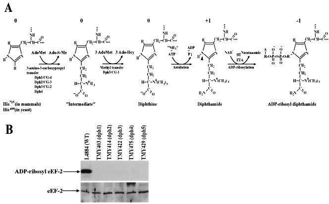
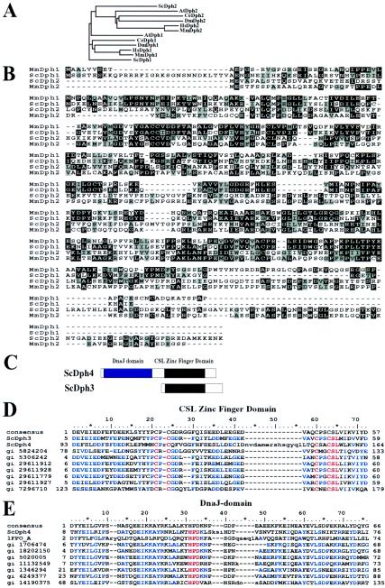
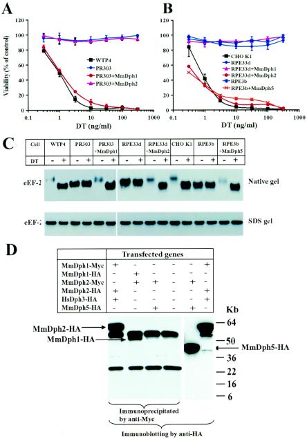
Diphthamide, a posttranslational modification of translation elongation factor 2 that is conserved in all eukaryotes and archaebacteria and is the target of diphtheria toxin, is formed in yeast by the actions of five proteins, Dph1 to -5, and a still unidentified amidating enzyme. Dph2 and Dph5 were previously identified. Here, we report the identification of the remaining three yeast proteins (Dph1, -3, and -4) and show that all five Dph proteins have either functional (Dph1, -2, -3, and -5) or sequence (Dph4) homologs in mammals. We propose a unified nomenclature for these proteins (e.g., HsDph1 to -5 for the human proteins) and their genes based on the yeast nomenclature. We show that Dph1 and Dph2 are homologous in sequence but functionally independent. The human tumor suppressor gene OVCA1, previously identified as homologous to yeast DPH2, is shown to actually be HsDPH1. We show that HsDPH3 is the previously described human diphtheria toxin and Pseudomonas exotoxin A sensitivity required gene 1 and that DPH4 encodes a CSL zinc finger-containing DnaJ-like protein. Other features of these genes are also discussed. The physiological function of diphthamide and the basis of its ubiquity remain a mystery, but evidence is presented that Dph1 to -3 function in vivo as a protein complex in multiple cellular processes.


Figures

FIG. 1.
FIG. 1.
DPH1 to DPH5 genes are required for diphthamide biosynthesis. (A) Diphthamide biosynthesis and ADP ribosylation. N-1 (arrowhead) of the histidine imidazole ring of diphthamide is the site for ADP ribosylation by DT and ETA. Ado-S-Me, methylthioadenosine; Ado-Hcy, S-adenosylhomocysteine. In ADP-ribosyl diphthamide, A, adenine moiety; R, ribosyl moiety. The relative net charges of the modified His side chains are indicated above each species. (B) Precise deletions of each of DPH1 to DPH5 genes result in the yeast mutants being completely resistant to ADP ribosylation by DT. (Top) Yeast cell lysates were incubated with [adenylate-32P]NAD and DT at room temperature for 30 min. The reactions were terminated by boiling the lysates in SDS sample buffer, and the samples were analyzed by SDS-PAGE, followed by autoradiography. (Bottom) Deletions of each of the DPH1 to DPH5 genes do not affect overall expression of eEF-2 protein as evidenced by Western blot analysis using an anti-eEF-2 antibody (a gift of Angus Nairn, Rockefeller University).
FIG.2.
FIG.2.
Sequence alignments of Dph proteins. (A) Phylogenetic analysis of Dph1 and Dph2 from different species. At, Arabidopsis thaliana; Ce, Caenorhabditis elegans. (B) Sequence alignments of yeast and mouse Dph1 and Dph2 proteins. Identical residues are shaded in black, and similar residues are shaded in gray. Dashes represent gaps in the aligned sequences. (C) Schematic representation of domain arrangements of ScDph3 and ScDph4 proteins. (D) The consensus CSL zinc finger domain sequence from the NCBI conserved-domain database aligned with the amino-terminal region of ScDph3 (residues 5 to 59), the carboxyl-terminal region of ScDph4 (residues 93 to 164), and other related proteins. In the alignment, identical residues are in blue and the four cysteine residues and the highly conserved CSL zinc finger motif are in red. (E) The consensus Dna-J sequence from the NCBI conserved-domain database aligned with the N-terminal region of ScDph4 (residues 8 to 76) and other related proteins. In the alignment, identical residues are in blue and the J-domain signature HPD motif is in red.
FIG. 3.
FIG. 3.
Complementation of the diphthamide-deficient CHO mutant cells by transfection with the respective mouse DPH genes. (A) Expression of MmDph1 in CG-4 PR303 cells complements their DT-resistant phenotype. Representative purified clones of PR303 cells transfected with mouse DPH1 or mouse DPH2 were incubated with various concentrations of DT for 48 h, and MTT was added to determine cell viability. Optical density readings were normalized to values for cells that received no toxin to calculate percent viability. (B) Expression of MmDph2 in CG-3 RPE33d cells and MmDph5 in CG-1 RPE3b cells complements their DT-resistant phenotypes. Cytotoxicity of DT to representative clones of RPE33d cells transfected with mouse DPH1 or mouse DPH2 and to a representative clone of RPE3b cells transfected with mouse DPH5 were determined as in panel A. (C) The CHO cell lysates were incubated with or without DT at room temperature for 30 min and then analyzed by either native PAGE (top) or SDS-PAGE (bottom), followed by Western blotting using an antibody against a linear peptide of the carboxyl terminus of eEF-2. +, present; −, absent. (D) Dph1 and Dph2 can physically interact in vivo. Combinations of HA- or Myc-tagged MmDph1, MmDph2, HsDph3, and MmDph5 expression plasmids as indicated were transiently transfected into CHO K1 cells for 24 h. Cell lysates were prepared and immunoprecipitated using an agarose-conjugated anti-Myc antibody, followed by Western blotting using an anti-HA antibody. Expression of MmDph5-HA in MmDph2-Myc- and MmDph5-HA-transfected cells was evidenced by direct Western blot analysis of the cell lysate using the anti-HA antibody. The two bands common in lanes 1 to 4 are to the heavy and light immunoglobulin G chains.

References

    1. Baudin, A., O. Ozier-Kalogeropoulos, A. Denouel, F. Lacroute, and C. Cullin. 1993. A simple and efficient method for direct gene deletion in Saccharomyces cerevisiae. Nucleic Acids Res. 21:3329-3330. - PMC - PubMed
    1. Bruening, W., A. H. Prowse, D. C. Schultz, M. Holgado-Madruga, A. Wong, and A. K. Godwin. 1999. Expression of OVCA1, a candidate tumor suppressor, is reduced in tumors and inhibits growth of ovarian cancer cells. Cancer Res. 59:4973-4983. - PubMed
    1. Burns, N., B. Grimwade, P. B. Ross-Macdonald, E. Y. Choi, K. Finberg, G. S. Roeder, and M. Snyder. 1994. Large-scale analysis of gene expression, protein localization, and gene disruption in Saccharomyces cerevisiae. Genes Dev. 8:1087-1105. - PubMed
    1. Carroll, S. F., J. T. Barbieri, and R. J. Collier. 1988. Diphtheria toxin: purification and properties. Methods Enzymol. 165:68-76. - PubMed
    1. Chen, C. M., and R. R. Behringer. 2004. Ovca1 regulates cell proliferation, embryonic development, and tumorigenesis. Genes Dev. 18:320-332. - PMC - PubMed

MeSH terms