. 2004 Nov;24(21):9682-94.
doi: 10.1128/MCB.24.21.9682-9694.2004.

The Mre11 nuclease is not required for 5' to 3' resection at multiple HO-induced double-strand breaks

Affiliations

The Mre11 nuclease is not required for 5' to 3' resection at multiple HO-induced double-strand breaks

Bertrand Llorente et al. Mol Cell Biol. 2004 Nov.

Abstract

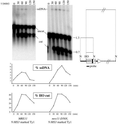
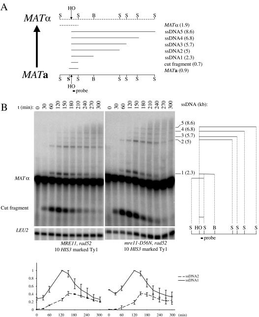
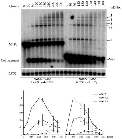
Current hypotheses suggest the Mre11 nuclease activity could be directly involved in double-strand break (DSB) resection in the presence of a large number of DSBs or limited to processing abnormal DNA ends. To distinguish between these possibilities, we used two methods to create large numbers of DSBs in Saccharomyces cerevisiae chromosomes, without introducing other substrates for the Mre11 nuclease. Multiple DSBs were created either by expressing the HO endonuclease in strains containing several HO cut sites embedded within randomly dispersed Ty1 elements or by phleomycin treatment. Analysis of resection by single-strand DNA formation in these systems showed no difference between strains containing MRE11 or the mre11-D56N nuclease defective allele, suggesting that the Mre11 nuclease is not involved in the extensive 5' to 3' resection of DSBs. We postulate that the ionizing radiation (IR) sensitivity of mre11 nuclease-defective mutants results from the accumulation of IR-induced DNA damage that is normally processed by the Mre11 nuclease. We also report that the processivity of 5' to 3' DSB resection and the yield of repaired products are affected by the number of DSBs in a dose-dependent manner. Finally, we show that the exonuclease Exo1 is involved in the processivity of 5' to 3' resection of an HO-induced DSB at the MAT locus.


Figures

FIG. 1.
FIG. 1.
Physical map of pBL001 containing the HOcs-marked Ty1 derived from the Ty1his3 AI (15). Striped arrowheads indicate Ty1 LTRs and the direction of Ty1 transcription. Open box, HIS3 gene (arrow, direction of its transcription); solid box, 104-bp AI (in antisense orientation relative to HIS3); vertical bars, relevant restriction sites (P, PvuII; N, NdeI; HO, HO cut site).
FIG. 2.
FIG. 2.
Single-stranded DNA intermediates are formed at the experimentally induced HO cut sites in MRE11 and mre11-D56N strains. DNA samples from MRE11 (LSY1264) (left) and mre11-D56N strains (LSY1299) (right) were prepared from cells undergoing a 60-min HO induction, digested with NdeI, electrophoresed under alkaline conditions, blotted onto a nylon membrane, and probed with a HIS3 fragment (see Materials and Methods). These two strains contain nine HIS3-marked Ty1s. Because splicing of the AI present in HIS3 is not an efficient process (15), some HOcs-HIS3 Ty1 elements still contain the 104-bp AI, as shown by the presence of two closely migrating bands at around 1.3 and 0.7-kb. t, time in minutes between the beginning of the HO induction and the collection of cells for DNA analysis; uncut, NdeI fragment from the HIS3-marked Ty1 containing the HO cleavage site; cut, HO-NdeI fragment overlapping with HIS3 from the HIS3-marked Ty1; ssDNA, ssDNA-containing fragment from the HO cleavage site of a particular HIS3-marked Ty1 and a downstream chromosomal NdeI cut site. (Bottom) Quantitation of the ssDNA intermediates and cut fragments for the two strains, expressed as percentages of their intensities relative to the sum of the intensities of all fragments at each time point. Symbols in the map to the right are as defined for Fig. 1.
FIG. 3.
FIG. 3.
In vivo HO cleavage of individual HO cut sites in a strain containing 10 HOcs-Ty1 elements (LSY1259). Left and right, 60- and 300-min HO induction, respectively. The arrows on the left start at the positions of the uncut parental PvuII fragments and end at the positions of the corresponding HO cut fragments (numbered 1 to 6 at the corresponding arrow). Any HO cut fragment is 1.6-kb smaller than its corresponding PvuII parental fragment. Stars indicate doublets, and the highest cut fragment comigrates with its uncut parental fragment (curved arrow). Two cut fragments comigrate with two of the smaller uncut parental fragments. As a result, only 6 cut fragments out of 10 are visible. Values are the percentages of the ratios of the intensities of the cut fragments to that of his3. t, time in minutes between the beginning of HO induction and the collection of cells for DNA analysis. Symbols in the map to the right are as defined for Fig. 1.
FIG. 4.
FIG. 4.
Visualization of GCRs by PFGE in a strain containing 10 marked Ty1s (LSY1259) after HO induction. The cells underwent a 60-min HO induction and were then plated on the noninducing medium. Three resulting individual colonies were used to prepare the DNA plugs (lanes 2 to 4). Lane 1 corresponds to the starting strain prior to HO induction. (Left) Pulsed-field gel stained with SYBR Gold. Arrows, chromosomes with an abnormal mobility; numbers on the left, chromosome numbers. (Right) Pulsed-field gel blotted and probed with HIS3. Numbers on the left, numbers of HIS3-marked Ty1s in the corresponding band in LSY1259; *, signal from the endogenous his3 gene.
FIG. 5.
FIG. 5.
5′ to 3′ resection at the MAT locus is unaffected by mre11-D56N in the presence of additional DSBs. (A) Physical map of the MATa- MATα switching process and illustration of the DNA intermediates as observed by alkaline gel electrophoresis after a StyI-BamHI double digestion. The probe used reveals the 0.9- and 1.9-kb StyI fragments from the uncut MATa and MATα loci, respectively. When expressed, HO cuts the MAT locus to produce a smaller 0.7-kb HO-StyI cut fragment. As a result of the 5′ to 3′ resection of the right end of the HO break, some StyI and BamHI cut sites become single stranded and resistant to cleavage, generating high-molecular-weight ssDNA fragments when electrophoresed under alkaline conditions (ssDNA1 to -5). The sizes of the different DNA intermediates are indicated in parentheses. S, StyI; B, BamHI; HO, HO cut site. (B) A 60-min HO induction was performed in MRE11 (LSY1477) and mre11-D56N (LSY1483) strains, and ssDNA formation at MAT was analyzed by alkaline gel electrophoresis (see Materials and Methods). The sizes of the ssDNA species (ssDNA1 to -5) are indicated in parentheses. LEU2, hybridization control; t, time in minutes between the beginning of HO induction and the collection of cells for DNA analysis. (Bottom) Quantitation of the ssDNA intensities, normalized with the LEU2 signal and expressed as the fraction of the maximum intensity of ssDNA1 for each strain. Error bars correspond to the range of the data from two independent experiments.
FIG. 6.
FIG. 6.
Decrease in ssDNA turnover after multiple HO-induced DSBs are made. A 60-min HO induction was performed in rad52 strains containing nine (LSY1481; left) or zero (LSY723; right) HIS3-marked Ty1s. (Top) ssDNA formation at MAT was analyzed by alkaline gel electrophoresis (see Materials and Methods). LEU2, hybridization control; t, time in minutes between the beginning of HO induction and the collection of cells for DNA analysis. (Bottom) Quantitation of the ssDNA intensities, normalized with the LEU2 signal and expressed as the fraction of the maximum intensity of ssDNA1 for each strain. Error bars correspond to the range of the data from two independent experiments.
FIG. 7.
FIG. 7.
Accumulation of ssDNA intermediates at MAT after phleomycin treatment. (Top) A 60-min HO induction was performed in two equivalent cultures of MRE11 (LSY678; left two panels) and mre11-D56N (LSY1032; right two panels) strains. Phleomycin was added to one culture of each strain during the entire period of HO induction, at a final concentration of 250 μg/ml (163 μM). ssDNA formation at MAT was analyzed by alkaline gel electrophoresis (see Materials and Methods). t, time in minutes between the beginning of HO induction and the harvest of the corresponding cells. Values for time zero for LSY678 with no phleomycin are not plotted because of partial DNA degradation. (Bottom) Quantitation of the ssDNA intensities, normalized with the LEU2 signal and expressed as the fraction of the maximum intensity of ssDNA1 for each strain. Error bars correspond to the range of the data from two independent experiments for mre11-D56N and the standard deviations of three independent experiments for MRE11.
FIG. 8.
FIG. 8.
Decreased resection of the HO-induced DSB at the MAT locus in exo1 mutants. Two 60-min HO inductions were performed in exo1 strains (LSY894-2B and LSY894-4C), and the formation of ssDNA intermediates generated was detected by alkaline gel electrophoresis. The amounts of ssDNA1 and ssDNA2 shown are expressed as the fractions of the maximum intensity of ssDNA1 for each strain. Error bars correspond to the range of the data from two independent experiments for exo1 and the standard deviations of three independent experiments for EXO1.

References

    1. Alani, E., R. Padmore, and N. Kleckner. 1990. Analysis of wild-type and rad50 mutants of yeast suggests an intimate relationship between meiotic chromosome synapsis and recombination. Cell 61:419-436. - PubMed
    1. Amundsen, S. K., and G. R. Smith. 2003. Interchangeable parts of the Escherichia coli recombination machinery. Cell 112:741-744. - PubMed
    1. Aylon, Y., and M. Kupiec. 2003. The checkpoint protein Rad24 of Saccharomyces cerevisiae is involved in processing double-strand break ends and in recombination partner choice. Mol. Cell. Biol. 23:6585-6596. - PMC - PubMed
    1. Aylon, Y., B. Liefshitz, G. Bitan-Banin, and M. Kupiec. 2003. Molecular dissection of mitotic recombination in the yeast Saccharomyces cerevisiae. Mol. Cell. Biol. 23:1403-1417. - PMC - PubMed
    1. Bartsch, S., L. E. Kang, and L. S. Symington. 2000. RAD51 is required for the repair of plasmid double-stranded DNA gaps from either plasmid or chromosomal templates. Mol. Cell. Biol. 20:1194-1205. - PMC - PubMed

Publication types

MeSH terms

LinkOut - more resources