. 2004 Nov;16(11):3033-44.
doi: 10.1105/tpc.104.025643. Epub 2004 Oct 14.

A novel molecular recognition motif necessary for targeting photoactivated phytochrome signaling to specific basic helix-loop-helix transcription factors

Affiliations

A novel molecular recognition motif necessary for targeting photoactivated phytochrome signaling to specific basic helix-loop-helix transcription factors

Rajnish Khanna et al. Plant Cell. 2004 Nov.

Abstract

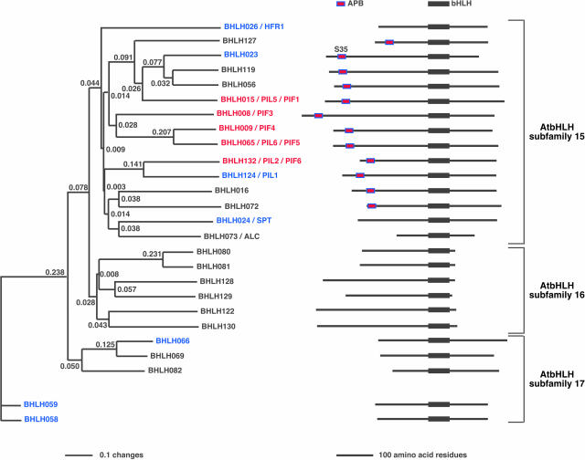
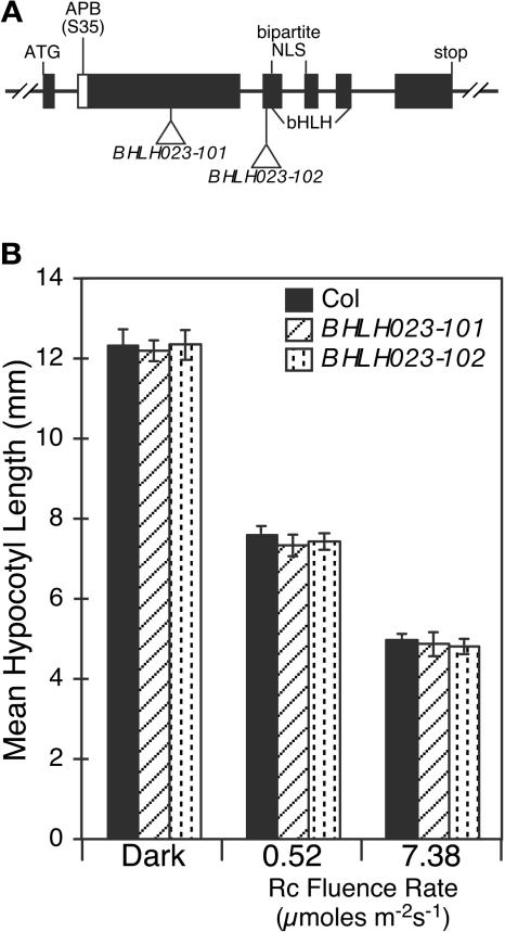
The phytochrome (phy) family of sensory photoreceptors (phyA to phyE) in Arabidopsis thaliana control plant developmental transitions in response to informational light signals throughout the life cycle. The photoactivated conformer of the photoreceptor Pfr has been shown to translocate into the nucleus where it induces changes in gene expression by an unknown mechanism. Here, we have identified two basic helix-loop-helix (bHLH) transcription factors, designated PHYTOCHROME-INTERACTING FACTOR5 (PIF5) and PIF6, which interact specifically with the Pfr form of phyB. These two factors cluster tightly with PIF3 and two other phy-interacting bHLH proteins in a phylogenetic subfamily within the large Arabidopsis bHLH (AtbHLH) family. We have identified a novel sequence motif (designated the active phytochrome binding [APB] motif) that is conserved in these phy-interacting AtbHLHs but not in other noninteractors. Using the isolated domain and site-directed mutagenesis, we have shown that this motif is both necessary and sufficient for binding to phyB. Transgenic expression of the native APB-containing AtbHLH protein, PIF4, in a pif4 null mutant, rescued the photoresponse defect in this mutant, whereas mutated PIF4 constructs with site-directed substitutions in conserved APB residues did not. These data indicate that the APB motif is necessary for PIF4 function in light-regulated seedling development and suggest that conformer-specific binding of phyB to PIF4 via the APB motif is necessary for this function in vivo. Binding assays with the isolated APB domain detected interaction with phyB, but none of the other four Arabidopsis phys. Collectively, the data suggest that the APB domain provides a phyB-specific recognition module within the AtbHLH family, thereby conferring photoreceptor target specificity on a subset of these transcription factors and, thus, the potential for selective signal channeling to segments of the transcriptional network.


Figures

Figure 1.
Figure 1.
PIF5 and PIF6 Bind Specifically to phyB (Pfr), whereas Six Other AtbHLH Proteins Tested Do Not Bind phyB. GAL4 activation domain (GAD) fusions (at N- or C-terminal domains) of full-length AtbHLH proteins were used as baits in in vitro coimmunoprecipitation assays. PhyB (Pfr) or phyB (Pfr) reconverted to Pr by a far-red light pulse (marked as Pr) were used as prey. Schematic diagram on the left shows the design of the experiments, and the SDS-PAGE separations of the pellet fractions are shown. (A) GAD:PIF3, PIF4:GAD, GAD:PIF5, and GAD:PIF6 specifically bind phyB (Pfr). GAD alone was used as control. (B) GAD:SPT, GAD:PIL1, GAD:BHLH023, GAD:BHLH058, GAD:BHLH059, and GAD:BHLH066 do not bind phyB. (C) Quantification of in vitro coimmunoprecipitation assays. The percentage of phyB recovered was calculated per bait. PhyB input is shown in the inset.
Figure 2.
Figure 2.
Members of the PIF3 Group of AtbHLH Proteins Contain a Conserved Sequence in their N-Terminal Domains. Neighbor-joining phylogenetic tree using full-length amino acid sequences of proteins closely related to PIF3. The tree was constructed with PAUP 4.0 software using an alignment (MultiAlin; Corpet, 1988) of predicted full-length amino acid sequences for each protein. All proteins are identified by their generic names and grouped in their subfamilies (Bailey et al., 2003), and other synonym/s are indicated. Twelve of the full-length AtbHLH proteins from different subfamilies have been tested for interactions with phyB using in vitro coimmunoprecipitation assays. Seven of the tested proteins did not interact (in blue) with phyB, whereas five show interactions (in red) specifically with the active Pfr form of phyB. Four AtbHLH proteins previously tested for interactions with phyB are HFR1 (Fairchild et al., 2000), PIF3 (Ni et al., 1998), PIF4 (Huq and Quail, 2002), and PIF1 (also known as PIL5; Yamashino et al., 2003; Huq et al., 2004). We propose two new names, PIF5 (old name, PIL6; Yamashino et al., 2003) and PIF6 (old name, PIL2; Yamashino et al., 2003) for the new phyB interacting factors to reflect their molecular activity. On the right, we show stick diagrams of the full-length proteins aligned at their bHLH domains. The presence of the APB consensus sequence in 12 AtbHLH proteins in subfamily 15 is indicated, including the S35 present in BHLH023, instead of the invariant G. The branch lengths are proportional to the indicated distance values (changes) between sequences. SPT (Heisler et al., 2001); ALC, ALCATRAZ (Rajani and Sundaresan, 2001). Proteins that interact with phyB (Pfr) are in red, and those that were tested but do not interact are in blue.
Figure 3.
Figure 3.
The APB Motif. (A) Alignments of the predicted N-terminal (1 to 100) amino acid sequences of 12 members of the AtbHLH subfamily 15 containing amino acid sequence homologies to the APB motif. Conserved amino acid sequences (in red) of the APB motif (blue outlined) are shown. The invariant amino acid residues required for APB function (reverse red font) are E31, L32, G37, and Q38 (PIF3 and PIF5 positions). The APB consensus is in bold type. (B) Phylogenetic neighbor-joining tree of the aligned APB sequences showing the putative evolutionary relationships of the APB motif between the family members. The distance values (changes) between sequences are proportional to the branch lengths (indicated). Proteins that interact with phyB (Pfr) are in red, and those that were tested but do not interact are in blue. The APB region of BHLH023 has a variation (S35 instead of invariant G).
Figure 4.
Figure 4.
The APB Motif Is Necessary for Binding phyB (Pfr). GAD fusions with full-length PIFs (containing wild-type APB, closed rectangles, or mutated APB, open rectangles) were used as baits in in vitro coimmunoprecipitation assays. PhyB (Pfr) or phyB (Pfr) reconverted to Pr by a far-red light pulse (marked as Pr) were used as prey. Schematic diagrams on the left show the design of the experiments, and the SDS-PAGE separations of the pellet fractions are shown on the right of each panel. (A) GAD:PIF5 binds phyB (Pfr) but the point mutations (E31A, L32A, G37A, and Q38A) abrogate binding to phyB. (B) APB mutations in PIF4:GAD (G35A) and GAD:PIF3 (E31A/G37A) eliminate their interactions with phyB (Pfr).
Figure 5.
Figure 5.
APB Motif Is Sufficient for Binding phyB (Pfr). (A) GAD:APB fusion proteins used as baits are sufficient for interactions with phyB (Pfr). GAD fusions with N-terminal (1 to 100) amino acid sequences containing the wild-type APB (closed rectangles) from PIF3, PIF4, PIF6, PIL1, and mutated APB (open rectangles) from PIF3 (E31A/G37A), PIF4 (G35A), and PIF6 (E19A/G25A) were tested for interactions with phyB. (B) Quantification of in vitro coimmunoprecipitation assays. PhyB input is shown in the inset. (C) The APB motif of PIF3 (GAD:APB) was tested for interactions with phyA, phyB, phyC, phyD, and phyE. The prey input is shown in the left panels.
Figure 6.
Figure 6.
APB Motif Activity Is Necessary for PIF4 Function. The srl2 mutant lines transformed with the PIF4:PIF4 full-length gene containing either wild-type APB [shown in red; PIF4 FL(1) and PIF4 FL(2)] or mutated APB [G35A shown in blue; PIF4 FL(G35A-1), PIF4 FL(G35A-2), and PIF4 FL(G35A-3)]. (A) Stick diagram showing the PIF4 promoter (2 kb upstream of the ATG) and the PIF4 gene used for transformations of srl2 mutant lines (position of srl2 T-DNA insertion in relation to the NLS and the bHLH is indicated). (B) Fluence rate response curves of mean hypocotyl lengths of wild-type (Ws), srl2 mutant, and indicated transgenic lines grown in Rc for 3 d. The phyB (Col) was used as a control. (C) Seedling photomorphogenic phenotypes of srl2 mutant seedlings grown in Rc (17.8 μmol m−2 s−1) for 3 d are rescued by the PIF4 FL(2) transgene, are overcompensated in PIF4 overexpressing transgenic line PIF4 FL(1), and are not rescued in PIF4 FL(G35A) transgenic lines. (D) Measurements of cotyledon area expansion in transgenic seedlings. The srl2 mutant seedlings grown for 3 d under Rc (17.8 μmol m−2 s−1) have enlarged cotyledon areas compared with the Ws (wild type), and this response is rescued (cotyledon expansion similar to the wild type) in the PIF4 FL(2) transgenic seedlings. The PIF4 FL(1) overexpressors show reduced cotyledon expansion; by contrast, all of the lines expressing mutated APB (G35A) have enlarged cotyledons compared with Ws. PhyB (Col) is shown for comparison. Approximately 20 to 25 seedlings for each line were used for measurements (40 to 50 cotyledons), and the values were normalized to Ws.
Figure 7.
Figure 7.
Relative PIF4 Transcript Levels Compared with Percentage of Mutant Phenotype Rescue in Transgenic Seedlings. (A) RNA gel blots showing Rc-induced expression of PIF4 message in transformants under control of the native PIF4 promoter. The Rc fold-induction values are indicated. All transgenes show approximately twofold induction in light, except for much higher levels detected in PIF4 FL(1). (B) Relative PIF4 transcript levels in Rc (top graph), normalized to the Ws levels, are shown with the percentage of recovery toward the wild-type phenotype (bottom graph; percentage of recovery is the percentage of mutant hypocotyl length rescue in transgenic lines). (C) Correlation between percentage of recovery (y axis) and relative PIF4 transcript levels (x axis) derived from the data presented above (B) for the indicated transgenic lines. Relative transcript level and percentage of recovery in the PIF4 FL transgenic lines (closed circles) and PIF4 FL(G35A) transgenic lines (open circles).
Figure 8.
Figure 8.
AtbHLH023 Mutant Seedlings Do Not Show Detectable Seedling Photomorphogenic Phenotypes. (A) Stick diagram showing the BHLH023 gene structure. Positions of the T-DNA insertions in BHLH023-101 and BHLH023-102 are indicated relative to the NLS and the bHLH domain. Based on the points of these insertions, the mutants would be predicted to be null for nuclear translocation and DNA binding if a stable truncated protein is made. The natural variation in the APB motif (S35) of BHLH023 is indicated. (B) Mean hypocotyl lengths of wild-type (Col), BHLH023-101, and BHLH023-102 mutant seedlings grown in Rc (0.52 or 7.38 μmol m−2 s−1) for 3 d.

Similar articles

Cited by

References

    1. Bailey, P.C., Martin, C., Toledo-Ortiz, G., Quail, P.H., Huq, E., Heim, M.A., Jakoby, J., Werber, M., and Weisshaar, B. (2003). Update on the basic helix-loop-helix transcription factor gene family in Arabidopsis thaliana. Plant Cell 15, 2497–2502. - PMC - PubMed
    1. Bauer, D., Viczián, A., Kircher, S., Nobis, T., Nitschke, R., Kunkel, T., Panigrahi, K., Ádám, E., Fejes, E., Schäfer, E., and Nagy, F. (2004). COP1 and multiple photoreceptors control degradation of PIF3, a transcription factor, required for light signalling in Arabidopsis. Plant Cell 16, 1433–1445. - PMC - PubMed
    1. Clough, S.J., and Bent, A.F. (1998). Floral dip: A simplified method for Agrobacterium-mediated transformation of Arabidopsis thaliana. Plant J. 16, 735–743. - PubMed
    1. Corpet, F. (1988). Multiple sequence alignment with hierarchical clustering. Nucleic Acids Res. 16, 10881–10890. - PMC - PubMed
    1. Fairchild, C.D., Schumaker, M.A., and Quail, P.H. (2000). HFR1 encodes an atypical bHLH protein that acts in phytochrome A signal transduction. Genes Dev. 14, 2371–2399. - PMC - PubMed

Publication types

MeSH terms