

Massive pulmonary artery thrombosis with haemoptysis in adults with Eisenmenger's syndrome: a clinical dilemma
- PMID: 15486107
- PMCID: PMC1768531
- DOI: 10.1136/hrt.2004.039198
Massive pulmonary artery thrombosis with haemoptysis in adults with Eisenmenger's syndrome: a clinical dilemma
Abstract
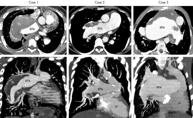
Although the frequency of haemoptysis in Eisenmenger's syndrome is well recognised, the high prevalence of pulmonary artery thrombus has been newly appreciated through the growing use of non-invasive imaging. Three patients with Eisenmenger's syndrome with haemoptysis are reported who underwent computed tomography pulmonary angiography and cardiovascular magnetic resonance. Each patient was found to have aneurysmal dilatation of the right pulmonary artery with large laminar thrombus. These cases illustrate a rising clinical problem in this special population-that is, how to treat and prevent large pulmonary artery thrombosis in the setting of haemoptysis. The authors discuss their approach to these cases and the known literature.
Figures

References
-
- Daliento L , Somerville J, Presbitero P, et al. Eisenmenger syndrome: factors relating to deterioration and death. Eur Heart J 1998;19:1845–55. - PubMed
-
- Canada WJ, Goodale F Jr, Currens JH. Defect of the interatrial septum, with thrombosis of the pulmonary artery; report of three cases. N Engl J Med 1953;248:309–16. - PubMed
-
- Perloff JK, Hart EM, Greaves SM, et al. Proximal pulmonary arterial and intrapulmonary radiologic features of Eisenmenger syndrome and primary pulmonary hypertension. Am J Cardiol 2003;92:182–7. - PubMed
-
- Silversides CK, Granton JT, Konen E, et al. Pulmonary thrombosis in adults with Eisenmenger syndrome. J Am Coll Cardiol 2003;42:1982–7. - PubMed
Publication types
MeSH terms
LinkOut - more resources
Full Text Sources
Medical