Case Reports
. 2004 Nov;90(11):1320.
doi: 10.1136/hrt.2004.035451.

Left ventricular diverticulum in a neonate with Cantrell syndrome

Affiliations
Case Reports

Left ventricular diverticulum in a neonate with Cantrell syndrome

S Di Bernardo et al. Heart. 2004 Nov.
No abstract available


Figures

Figure 1
Figure 1
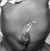
Thorax and abdomen demonstrating the abdominal mass on the midline, being the left ventricular diverticulum (LVD), as well as the small omphalocoele (OP).
Figure 2
Figure 2
Angiography of the left ventricle (LV) with the catheter coming from the vena cava inferior through the foramen ovale, demonstrating the dextroverse position of the heart and the left ventricular diverticulum (LVD), passing through the diaphragm pointing at the umbilicus. RCA, right coronary artery; AO, ascending aorta.
Figure 3
Figure 3
Anatomical view during surgery with a sternotomy and a midline laparotomy revealing the dextroverse position of the heart, and the left ventricular diverticulum (LVD) prolonging the left ventricle (LV) into the abdomen through the diaphragmatic hernia and terminating at the umbilicus in the omphalocoele. RV, right ventricle; PA, main pulmonary artery.

Publication types