. 2004 Oct 14;32(18):5553-69.
doi: 10.1093/nar/gkh876. Print 2004.

Protein kinase CK2 phosphorylation regulates the interaction of Kaposi's sarcoma-associated herpesvirus regulatory protein ORF57 with its multifunctional partner hnRNP K

Affiliations

Protein kinase CK2 phosphorylation regulates the interaction of Kaposi's sarcoma-associated herpesvirus regulatory protein ORF57 with its multifunctional partner hnRNP K

Poonam Malik et al. Nucleic Acids Res. .

Retraction in

Expression of concern in

Abstract

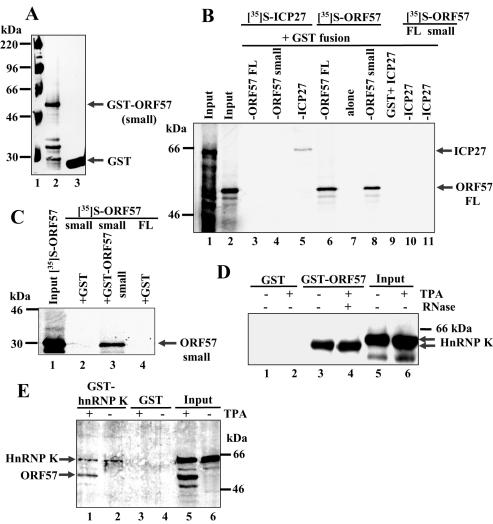
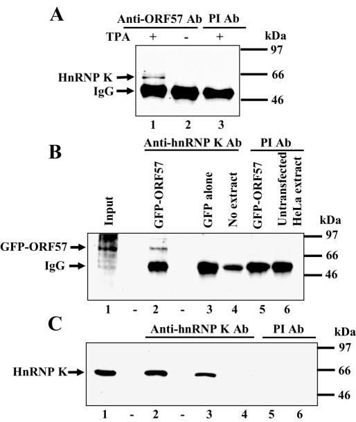
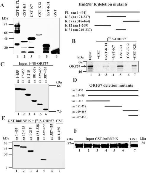
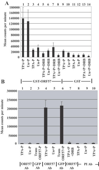
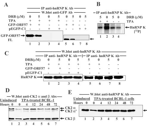
ORF57 protein of Kaposi's sarcoma-associated herpesvirus has a counterpart in all herpesvirus of mammals and birds and regulates gene expression at transcriptional and post-transcriptional levels. ORF57 was capable of self-interaction and bound a rapidly migrating form of heterogeneous nuclear ribonucleoprotein K (hnRNP K), a multifunctional cellular protein involved in gene expression. In virus infected cell extracts, ORF57 was present in a complex with hnRNP K that had protein kinase CK2 activity, and was phosphorylated by CK2. Different regions of ORF57 bound both catalytic alpha/alpha' and regulatory beta subunits of CK2. CK2 modification enhanced the ORF57-hnRNP K interaction, and may regulate the presence and activities of components in the complex. We suggest that ORF57 and hnRNP K interaction may modulate ORF57-mediated regulation of viral gene expression. Herpesviral ORF57 (Rhadinovirus) and ICP27 (Simplexvirus) proteins both interact with hnRNP K and CK2 implying that adaptation of the ancestral hnRNP K and CK2 to associate with viral regulatory ancestor protein likely pre-dates divergence of these Herpesviridae genera that occurred 200 million years ago.


Figures

Figure 1
Figure 1
ORF57 protein is capable of self-interaction and interacts with hnRNP K from KSHV-infected cell extracts using pull down assays. (A) Expression of recombinant GST–ORF57 small encoding ORF57 amino acids 181–455. GST–ORF57 small (lane 2) and GST alone (lane 3) separated by SDS–PAGE and visualized by Coomassie blue staining. (B) ORF57 protein interacts with itself but not with its HSV-1 ICP27 homologue. The pull down assay was performed with GST–ORF57 FL and small, GST–ICP27 or GST alone and [35S]-labelled ORF57 FL or small or ICP27 proteins using the following: input labelled ICP27 (lane 1); input labelled ORF57 FL (lane 2); GST–ORF57 FL + labelled ICP27 (lane 3); GST–ORF57 small + labelled ICP27 (lane 4); GST–ICP27 + labelled ICP27 (lane 5); GST–ORF57 FL + labelled ORF57 FL (lane 6); GST + labelled ORF57 FL (lane 7); GST–ORF57 small + labelled ORF57 FL (lane 8); GST + labelled ICP27 (lane 9); GST–ICP27 + labelled ORF57 FL (lane 10); GST–ICP27 + labelled ORF57 small (lane 11). (C) ORF57 self-interaction does not involve N-terminal amino acids 1–180. Pull down assays were followed by phosphorimager analysis using the following: input labelled ORF57 small (amino acids 181–455) (lane 1); GST + labelled ORF57 small (lane 2); GST–ORF57 small + labelled ORF57 small (lane 3); GST + labelled ORF57 FL (lane 4). (D) A rapidly migrating hnRNP K form from TPA-treated and untreated BCBL-1 cells is pulled down by GST–ORF57. TPA-treated and untreated BCBL-1 cell extracts were used in pull down assays, bound proteins were eluted and resolved by SDS–PAGE then immunoblotted with anti-hnRNP K Ab using the following: untreated extract + GST (lane 1); TPA-treated extract + GST (lane 2); untreated extract + GST–ORF57 FL (lane 3); TPA-treated extract + GST–ORF57 FL + RNAse (lane 4); input untreated extract (lane 5); input TPA-treated extract (lane 6). (E) GST–hnRNP K pulls down ORF57 and hnRNP K proteins from TPA-treated cells. After pull down assays using BCBL-1 cell extracts, bound proteins fractionated by SDS–PAGE were subjected to western blotting with a mixture of anti-ORF57 and anti-hnRNP K rabbit Abs using the following: TPA-treated extract + GST–hnRNP K (lane 1); untreated extract + GST–hnRNP K (lane 2); TPA-treated extract + GST (lane 3); untreated extract + GST (lane 4); input TPA-treated extract (lane 5); input untreated extract (lane 6).
Figure 2
Figure 2
ORF57 protein interacts with hnRNP K using immunoprecipitation assays. (A) ORF57 and hnRNP K proteins are co-immunoprecipitated from KSHV-infected cell extracts. BCBL-1 cell extracts were treated with anti-ORF57 Abs or pre-immune Ab, immunoprecipitates were separated by SDS–PAGE, then gels were western blotted with anti-hnRNP K Ab using the following: TPA-treated extract + anti-ORF57 Ab (lane 1); untreated extract + anti-ORF57 Ab (lane 2); TPA-treated extract + pre-immune Ab (lane 3). The strong band of 50 kDa represents IgG heavy chains of the precipitating Ab. (B) ORF57 protein co-immunoprecipitated with anti-hnRNP K Ab from HeLa cells transfected with an ORF57 expression vector. Immunoprecipitations were performed on extracts of HeLa cells transfected with plasmids pEGFP-ORF57 FL or pEGFP-C1 empty vector using anti-hnRNP K Ab or pre-immune Ab. Immunoprecipitates were separated by SDS–PAGE, then gels were western blotted using anti-GFP Ab with the following: input pEGFP-ORF57 extract (lane 1); pEGFP-ORF57 transfected cell extract + anti-hnRNP K Ab (lane 2); pEGFP-C1 transfected extract + anti-hnRNP K Ab (lane 3); no extract, only protein A-agarose beads + anti-hnRNP K Ab (lane 4); pEGFP-ORF57 transfected extract + pre-immune Ab (lane 5); untransfected extracts + pre-immune Ab (lane 6). (C) HnRNP K immunoprecipitates from both transfected and untransfected HeLa cell extracts. Western blotting the immunoprecipitates shown in Figure 2B using anti-hnRNP K Ab indicated that hnRNP K was co-immunoprecipitated in the appropriate samples.
Figure 3
Figure 3
Mapping the regions of hnRNP K and ORF57 proteins required for their interaction. (A) Left panel: GST–hnRNP K FL (amino acids 1–464) or four deletion mutants were used in the pull down assay with [35S]-labelled ORF57 FL. GST–hnRNP K FL and deletion mutant proteins were separated by SDS–PAGE and visualized by Coomassie blue staining as follows: GST–hnRNP K FL (lane 1); GST–hnRNP K 3 amino acids 171–337 (lane 2); GST–hnRNP K 7 amino acids 318–464 (lane 3); GST–hnRNP K 12 amino acids 1–209 (lane 4); GST–hnRNP K 31 amino acids 240–337 (lane 5); GST alone (lane 6). Right panel: cartoon showing GST–hnRNP K FL and the various deletion mutants used. (B) Mapping regions of hnRNP K that interacted with ORF57. Bound proteins were resolved by SDS–PAGE, gels were analysed by autoradiography using the following: input labelled ORF57 FL (lane 1); labelled ORF57 + GST (lane 2); labelled ORF57 + GST–hnRNP K FL (lane 3); labelled ORF57 + GST–hnRNP K 7 amino acids 318–464 (lane 4); labelled ORF57 + GST–hnRNP K 3 amino acids 171–337 (lane 5); labelled ORF57 + GST–hnRNP K 12 amino acids 1–209 (lane 6); labelled ORF57 + GST–hnRNP K 31 amino acids 240–337 (lane 7). (C) Expression of [35S]-labelled ORF57 deletion mutant proteins used in mapping as follows: ORF57 FL amino acids 1–455 (lane 1); ORF57 amino acids 17–455 (lane 2); amino acids 1–215 (lane 3); amino acids 181–328 (lane 4); amino acids 329–455 (lane 5); amino acids 387–455 (lane 6). (D) Cartoon showing ORF57 FL and the various ORF57 deletion mutants used. (E) Mapping regions of ORF57 that interacted with hnRNP K. GST–hnRNP K FL was used in pull down assays with labelled ORF57 proteins, bound proteins were separated by SDS–PAGE and gels analysed by phosphorimaging using the following: labelled ORF57 amino acids 1–455 (lane 1); labelled ORF57 amino acids 17–455 (lane 2); labelled ORF57 amino acids 1–215 (lane 3); labelled ORF57 amino acids 181–328 (lane 4); labelled ORF57 amino acids 329–455 (lane 5); labelled ORF57 amino acids 387–455 (lane 6); GST alone + labelled ORF57 FL (lane 7). (F) Use of similar amounts of GST–hnRNP K FL in the pull down assays shown in Figure 3E was demonstrated by western blotting similar amounts of samples eluted from the pull downs using anti-GST Ab.
Figure 4
Figure 4
Protein kinase CK2 activity is present in complexes containing ORF57 and hnRNP K proteins. A sensitive and specific method to determine CK2 activity in immunoprecipitates uses an artificial peptide substrate (25). Error bars show the mean values (± SD) obtained using assays from three independent experiments performed in duplicate. (A) CK2 activity is present in complexes containing ORF57 protein. The pull down assay using GST–ORF57 FL (bars 1–8) or GST alone (bars 9–14) with extracts of BCBL-1 cells was followed by a CK2 activity assay. CK2 assays used TPA-treated (TPA) and untreated (Un) cell extracts, and were performed with (+P) or without (−P) peptide and in the presence (+DRB) or absence of 50 μM DRB, a specific CK2 inhibitor, that acts in vitro and in vivo. (B) Extracts of TPA-treated or untreated BCBL-1 cells and also transfected with pEGFP-gORF57 FL (Trans ORF57) were immunoprecipitated with anti-ORF57 Ab (bars 1, 2, 4, 5, and 7), anti-GFP Ab (bars 3 and 6) or pre-immune (PI) serum Ab (bars 8–10), and CK2 activity present in the immunoprecipitates was assayed. (C) CK2 activity is present in complexes containing hnRNP K and ORF57 proteins. CK2 activities present in immunoprecipitates generated by anti-hnRNP K Ab from extracts of TPA-treated or untreated BCBL-1 cells. Bars 1–5, immunoprecipitations with anti-hnRNP K Ab; bars 6 and 7, immunoprecipitations with pre-immune serum Ab. (D) CK2 activities present in immunoprecipitates generated from 293 cell extracts following transfection with expression plasmids, pEGFP-ORF57 small, pEGFP-gORF57 FL and pcDNA3-gORF50. Immunoprecipitations with anti-hnRNP K Ab (bars 1–10); immunoprecipitations with pre-immune serum Ab (bars 11–13); CK2 activities in the presence of DRB (bars 3, 6, and 13).
Figure 4
Figure 4
Protein kinase CK2 activity is present in complexes containing ORF57 and hnRNP K proteins. A sensitive and specific method to determine CK2 activity in immunoprecipitates uses an artificial peptide substrate (25). Error bars show the mean values (± SD) obtained using assays from three independent experiments performed in duplicate. (A) CK2 activity is present in complexes containing ORF57 protein. The pull down assay using GST–ORF57 FL (bars 1–8) or GST alone (bars 9–14) with extracts of BCBL-1 cells was followed by a CK2 activity assay. CK2 assays used TPA-treated (TPA) and untreated (Un) cell extracts, and were performed with (+P) or without (−P) peptide and in the presence (+DRB) or absence of 50 μM DRB, a specific CK2 inhibitor, that acts in vitro and in vivo. (B) Extracts of TPA-treated or untreated BCBL-1 cells and also transfected with pEGFP-gORF57 FL (Trans ORF57) were immunoprecipitated with anti-ORF57 Ab (bars 1, 2, 4, 5, and 7), anti-GFP Ab (bars 3 and 6) or pre-immune (PI) serum Ab (bars 8–10), and CK2 activity present in the immunoprecipitates was assayed. (C) CK2 activity is present in complexes containing hnRNP K and ORF57 proteins. CK2 activities present in immunoprecipitates generated by anti-hnRNP K Ab from extracts of TPA-treated or untreated BCBL-1 cells. Bars 1–5, immunoprecipitations with anti-hnRNP K Ab; bars 6 and 7, immunoprecipitations with pre-immune serum Ab. (D) CK2 activities present in immunoprecipitates generated from 293 cell extracts following transfection with expression plasmids, pEGFP-ORF57 small, pEGFP-gORF57 FL and pcDNA3-gORF50. Immunoprecipitations with anti-hnRNP K Ab (bars 1–10); immunoprecipitations with pre-immune serum Ab (bars 11–13); CK2 activities in the presence of DRB (bars 3, 6, and 13).
Figure 5
Figure 5
ORF57 protein interacts with CK2 α, α′ and β subunits. (A) The recombinant proteins used were visualized by Coomassie blue staining as follows: molecular weight marker (lane1); GST–CK2 α FL (lane 2); GST–CK2 α′ FL (lane 3); GST alone (lane 4); MBP alone (lane 5); MBP–CK2 β FL (lane 6). (B) The pull down assay was performed using [35S]-labelled ORF57 FL, small or luciferase proteins with recombinant CK2 subunit fusion proteins. Following sample separation by SDS–PAGE, gels were analysed by autoradiography using the following: input labelled ORF57 FL (lane 1); input labelled ORF57 small (lane 2); GST–CK2 α + ORF57 FL (lane 3); GST–CK2 α + ORF57 small (lane 4); GST–CK2 α′ + ORF57 FL (lane 5); GST–CK2 α′ + ORF57 small (lane 6); MBP–CK2 β + ORF57 FL (lane 7); MBP–CK2 β + ORF57 small (lane 8); GST alone + ORF57 FL (lane 9); MBP alone + ORF57 FL (lane 10); GST–CK2 α + luciferase (lane 11). (C) The pull down assay was performed using TPA-treated or untreated BCBL-1 cell extracts. Following sample separation by SDS–PAGE, gels were western blotted with anti-ORF57 Ab using the following: input untreated extract (lane 1); input TPA-treated extract (lane 2); GST–CK2 α + TPA-treated cells (lane 3); GST–CK2 α + untreated cells (lane 4); GST–CK2 α′ + TPA-treated cells (lane 5); GST–CK2 α′ + untreated cells (lane 6); GST alone + TPA-treated cells (lane 7); MBP alone + TPA-treated cells (lane 8); MBP–CK2 β + untreated cells (lane 9); MBP–CK2 β + TPA-treated cells (lane 10). The band above the ORF57 protein (Figure 5C, lanes 9 and 10, marked with asterisk) is an MBP–CK2 β species that cross-reacts with anti-ORF57 Ab present in both TPA-treated and untreated cell extracts. (D) The pull down assay was performed with ORF57 and CK2 subunits, all recombinant proteins expressed from prokaryotic vectors in Escherichia coli. GST–ORF57 FL or GST alone bound to glutathione beads were mixed with equivalent amounts of recombinant His–CK2 α, or MBP–CK2 β, or His–thioredoxin fusion proteins. Following sample separation by SDS–PAGE, gels western blotted using a mixture of anti-CK2 α and β Abs (Figure 5D, lanes 1–4) or with anti-His Ab (Figure 5D, lane 5) using the following: GST–ORF57 FL + His–CK2 α (lane 1); GST alone + His–CK2 α (lane 2); GST–ORF57 FL + MBP–CK2 β (lane 3); GST alone + MBP–CK2 β (lane 4); GST–ORF57 FL + His–thioredoxin (lane 5).
Figure 6
Figure 6
Mapping the ORF57 regions involved in interaction with CK2 α/α′ and β subunits protein and vice versa. (A–D) ORF57 regions required for interaction with CK2 α′ and CK2 β were mapped using pull down assays with GST–CK2 α′ or GST alone, and MBP–CK2 β or MBP alone, with [35S]-labelled ORF57 FL and its deletion mutants. (A) The pull down assay was performed with GST–CK2 α′ and different labelled ORF57 truncations using the following: ORF57 FL amino acids 1–455 (lane 1); ORF57 small amino acids 181–455 (lane 2); ORF57 amino acids 1–215 (lane 3); ORF57 amino acids 181–328 (lane 4); ORF57 amino acids 329–455 (lane 5); ORF57 amino acids 387–455 (lane 6); GST alone + ORF57 FL (lane 7). (B) The pull down assay was performed with MBP–CK2 β and different labelled ORF57 truncations using the following: ORF57 FL (lane 1); ORF57 amino acids 17–455 (lane 2); ORF57 amino acids 1–215 (lane 3); ORF57 amino acids 181–328 (lane 4); ORF57 amino acids 329–455 (lane 5); ORF57 amino acids 387–455 (lane 6); MBP alone + ORF57 FL (lane 7). (C) Equal portions of the samples shown in Figure 6A examined by western blotting using anti-GST Ab. (D) Equal portions of the samples shown in Figure 6B examined by western blotting using anti-MBP Ab. (E) Mapping the CK2 β regions that interact with [35S]-labelled ORF57. Phosphorimager analysis using the following: input ORF57 FL (lane 1); MBP–CK2 β FL + ORF57 FL + RNase (lane 2); MBP–CK2 β amino acids 1–182 + ORF57 FL (lane 3); MBP–CK2 β amino acids 1–110 + ORF57 FL (lane 4); MBP alone + ORF57 FL (lane 5); MBP–CK2 β FL + ORF57 small amino acids 181–455 (lane 6); GST–CK2 β amino acids 1–150 + ORF57 FL (lane 7); GST–CK2 β amino acids 51–150 + ORF57 FL (lane 8); GST–CK2 β amino acids 1–55 + ORF57 FL (lane 9); GST alone + ORF57 FL (lane 10); MBP–CK2 β FL + luciferase (lane 11).
Figure 7
Figure 7
Phosphorylation of ORF57 by CK2 in vitro and in KSHV-infected cells. (A) The ability of CK2 to phosphorylate GST–ORF57 FL in vitro was examined by mixing beads carrying ORF57 protein with recombinant CK2 holoenzyme and phosphorylation reactions were carried out in the presence or absence of 50 μM DRB. Proteins were separated by SDS–PAGE and gels exposed to a phosphorimager screen using the following: CK2 holoenzyme + GST–ORF57 FL (lane 1); CK2 holoenzyme + GST–ORF57 FL + DRB (lane 2). (B) Use of equivalent amounts of GST–ORF57 was determined by western blotting portions of the phosphorylation reactions shown in Figure 7A with anti-ORF57 (GH) Ab. (C) To examine in vivo phosphorylation of ORF57 by CK2, BCBL-1 cells were TPA-treated or left untreated and labelled with [35S]-methionine or [32P]-orthophosphate in presence or absence of DRB. Immunoprecipitates obtained using anti-ORF57 Ab were separated by SDS–PAGE, and gels were exposed to a phosphorimager screen using the following: Untreated cells + [35S]-methionine (lane 1); TPA-treated cells + [35S]-methionine (lane 2); untreated cells + [32P]-orthophosphate (lane 3); TPA-treated cells + [32P]-orthophosphate (lane 4); TPA-treated cells + [32P]-orthophosphate + 10 μM DRB (lane 5); TPA-treated cells + [32P]-orthophosphate + 5 μM DRB (lane 6). (D) [32P]-orthophosphate labelled BCBL-1 cell extracts used in Figure 7C prior to immunoprecipitations were western blotted with anti-ORF57 (GH) peptide Ab to assess the levels of ORF57 protein using the following: untreated cells (lane 1); TPA-treated cells (lane 2); TPA-treated cells in presence of 10 μM DRB (lane 3); TPA-treated cells in presence of 5 μM DRB (lane 4).
Figure 8
Figure 8
Phosphorylation by CK2 enhances the interaction between ORF57 and hnRNP K proteins. (A) BCBL-1 cells were transfected with either pEGFP-gORF57 FL or pEGFP-C1, and with pCH110 encoding β-Gal as an internal control for transfection efficiency, and then were either treated with TPA (lanes 1–4) or left untreated (lanes 5–8). Immunoprecipitates of cell extracts obtained with anti-hnRNP K Ab were separated by SDS–PAGE and western blotted with anti-GFP Ab using the following: pEGFP-gORF57 FL (lanes 1 and 5); pEGFP-gORF57 FL + 5 μM DRB (lanes 2 and 6); pEGFP-C1 (lanes 3 and 7); pEGFP-C1 + 5 μM DRB (lanes 4 and 8). Position of GFP-ORF57 FL is indicated by an arrow. (B) HnRNP K protein is differentially phosphorylated by CK2 in TPA-treated as compared to untreated BCBL-1 cells. TPA-treated or untreated BCBL-1 cells in the presence or absence of 5 μM DRB were labelled with [32P]-orthophosphate for 6 h. Immunoprecipitates obtained using anti-hnRNP K Ab were separated by SDS–PAGE, followed by autoradiography using the following: TPA-treated (lanes 1 and 2); untreated cell extracts (lanes 3 and 4); in presence of DRB (lanes 1 and 3). (C) Western blotting using anti-hnRNP K Ab of anti-hnRNP K Ab immunoprecipitates used in Figure 8A from the TPA-treated transfected BCBL-1 cell extracts (lanes 1–4) and used in Figure 8B from [32P]-orthophosphate labelled cell extracts (lanes 5–8). (D and E) Extracts of BCBL-1 cells untreated or treated with TPA at various time points were western blotted with either a mixture of anti-CK2 α and β subunit Abs (Figure 8D) or anti-hnRNP K Ab (Figure 8E) using the following: untreated cells (lane 1); TPA-treated from 0–72 h (lanes 2–7). Approximately equal amounts of CK2 subunits or hnRNP K protein were present; IP, immunoprecipitation; W.blot, western blotting.

References

    1. Davison A.J. and Clements,J.B. (1997) Herpesviruses: general properties. In Mahy,B.W.J. and Collier,L.H. (eds), Topley & Wilson's Principles of Bacteriology, Virology and Immunology. 9th edn. Edward Arnold, London, pp. 309–323.
    1. Roizman B. and Knipe,D.M. (2001) In Knipe,D.M. and Howley,P.M. (eds), Fields Virology. 4th edn. Lippincott Williams & Wilkins, Philadelphia, Vol. 2, pp. 2399–2459.
    1. Chang Y., Cesarman,E., Pessin,M.S., Lee,F., Culpepper,J., Knowles,D.M. and Moore,P.S. (1994) Identification of herpesvirus-like DNA sequences in AIDS-associated Kaposi's sarcoma. Science, 266, 1865–1869. - PubMed
    1. Dourmishev L.A., Dourmishev,A.L., Palmeri,D., Schwartz,R.A. and Lukac,D.M. (2003) Molecular genetics of Kaposi's sarcoma-associated herpesvirus (human herpesvirus 8): epidemiology and pathogenesis. Microbiol. Mol. Biol. Rev., 67, 175–212. - PMC - PubMed
    1. Viejo-Borbolla A. and Schulz,T.F. (2003) Kaposi's sarcoma-associated herpesvirus (KSHV/HHV8): key aspects of epidemiology and pathogenesis. AIDS Rev., 5, 222–229. - PubMed

Publication types

MeSH terms