. 2004 Oct 14;32(18):5582-95.
doi: 10.1093/nar/gkh892. Print 2004.

The sequences and activities of RegB endoribonucleases of T4-related bacteriophages

Affiliations

The sequences and activities of RegB endoribonucleases of T4-related bacteriophages

Lina Piesiniene et al. Nucleic Acids Res. .

Abstract

The RegB endoribonuclease encoded by bacteriophage T4 is a unique sequence-specific nuclease that cleaves in the middle of GGAG or, in a few cases, GGAU tetranucleotides, preferentially those found in the Shine-Dalgarno regions of early phage mRNAs. In this study, we examined the primary structures and functional properties of RegB ribonucleases encoded by T4-related bacteriophages. We show that all but one of 36 phages tested harbor the regB gene homologues and the similar signals for transcriptional and post-transcriptional autogenous regulation of regB expression. Phage RB49 in addition to gpRegB utilizes Escherichia coli endoribonuclease E for the degradation of its transcripts for gene regB. The deduced primary structure of RegB proteins of 32 phages studied is almost identical to that of T4, while the sequences of RegB encoded by phages RB69, TuIa and RB49 show substantial divergence from their T4 counterpart. Functional studies using plasmid-phage systems indicate that RegB nucleases of phages T4, RB69, TuIa and RB49 exhibit different activity towards GGAG and GGAU motifs in the specific locations. We expect that the availability of the different phylogenetic variants of RegB may help to localize the amino acid determinants that contribute to the specificity and cleavage efficiency of this processing enzyme.


Figures
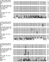
Figure 1
Figure 1
Amino acid sequence alignment of RegB endoribonucleases of T4-related phages. The RegB sequences of the phages were aligned with the T4 RegB sequence using the ClustalW program. A white background indicates amino acid motifs common to all of the T4-related subgroups. The sequences shown with black backgrounds are not well conserved. A dash indicates a space, which was inserted in the sequence to preserve the alignment. An asterisk (*) means that the residues in the column are identical; a double dot (:) indicates the conserved substitutions; a dot (.) indicates the semi-conserved substitutions.
Figure 2
Figure 2
Genes and promoters in the 1.10 kb genomic region with gene regB of bacteriophages T4, RB69 (TuIa) and RB49. The locations of early promoters are shown, as well as the positions of potential RegB cleavage sites within the SD sequences and in the coding sequences of regB. Vertical arrows show the sites susceptible to the RegB cleavage.
Figure 3
Figure 3
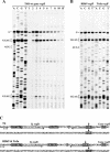
Primer extension analyses of transcripts for gene regB of phages T4, RB69 and TuIa. (A) Primer extension sequencing reaction of RNA isolated from E.coli BE cells at 5 min post-infection with bacteriophage T4 at 30°C. Primer extension reactions of RNA isolated at 1–15 min post-infection from the cells that were infected with phage T4 at 30°C are shown next to the sequencing reactions. (B) Primer extension sequencing reactions of RNA isolated from E.coli BE cells at 5 min post-infection with either phage RB69 or TuIa at 30°C. Primer extension sequencing reactions were performed using the primers Pr. T4regB(R1) (A) and Pr. RB69regB(R1) (B). The sequencing lanes are labeled with the dideoxynucleotides used in the sequencing reactions. The time (minutes) of post-infection that each RNA was isolated is noted at the top of the figures. The initiating nucleotides for the transcripts, the GGAG motifs within the SD sequences, the initiating codons, as well as the GGAG motifs in the coding region of gene regB mRNA are noted. (C) The nucleotide sequences of the 5′ flanking region of gene regB of phages T4, RB69 and TuIa. The −35 and −10 regions of the early promoters, the initiating nucleotides for the transcripts, the GGAG motifs within the SD sequences, the initiation codons, as well as the GGAG motifs in coding regions of genes, are shown with black backgrounds. Vertical arrows denote the positions of RegB cleavage. Convergent arrows indicate inverted repeats. Asterisks indicate the termination codons for the upstream genes.
Figure 4
Figure 4
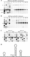
Primer extension analyses of phage RB49 transcript for gene regB. (A and B) Primer extension sequencing reactions of RNA isolated from E.coli BE cells at 5 min post-infection with RB49 at 30°C. Primer extension reactions of RNA isolated at 1–15 min post-infection from cells that were infected with RB49 at 30°C are shown next to the sequencing reactions. The sequencing lanes are labeled with dideoxynucleotides used in the sequencing reactions. The time (minutes) of post-infection that each RNA was isolated is noted at the top of the figures. (C) Primer extension sequencing of RNA isolated from E.coli N3433 (rne+) and N3431 (rne) cells at 5 min post-infection with RB49 at 30 and 43°C. The GGAU motif within the SD sequence, the initiation codon for regB gene, the initiating nucleotide for the RegB-dependent transcript, as well as the 5′ end nucleotide of RNase E processed transcript are noted. Primer extension sequencing reactions were performed using primers Pr. RB49regB(R1) (A) and Pr. RB49regB(R2) (B and C). (D) Nucleotide sequence flanking the RNase E cleavage site of the gene regB transcript and the potential secondary structures close to the cleavage site are shown. The arrow indicates the position of the RNase E cleavage. The 5′ end nucleotide of RNase E processed transcript is shown with a black background. Asterisks indicate the termination codon for gene regB.
Figure 5
Figure 5
Primer extension sequencing of the transcripts for genes regB.1 (A), regB (B), vs.1 (C) and 43 (D) of phage RB49. Primer extension sequencing reactions of RNA isolated at 5 min post-infection from E.coli BE cells that were infected with RB49 in the absence or in the presence of chloramphenicol at 30°C. Sequencing of DNA fragment carrying gene 43 of phage RB49 is also presented (D). The initiating nucleotides for the transcripts, the potential RegB cleavage sites within the SD sequences, as well as initiation codons for the corresponding genes are noted. Convergent arrow indicates inverted repeat sequences. Primer extension reactions were performed using primers Pr. RB49regB.1(R1) (A), Pr. RB49regB(R1) (B), Pr. RB49vs.1(R1) (C) and Pr. RB49g43(R1) (D). The nucleotide sequences of the 5′ flanking region of genes regB.1, regB, vs.1 and 43 of phage RB49 are shown at the bottom of the figure. The −35 and −10 regions of the early promoters, the initiating nucleotides for the transcripts, the GGAG and GGAU motifs within the SD sequences, as well as initiation codons are shown with black backgrounds. Vertical arrows denote the position of RegB cleavage. Convergent arrows indicate inverted repeats. Asterisks indicate the termination codons for the upstream genes.
Figure 6
Figure 6
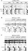
Susceptibility of RB49 transcripts for genes vs.1 and 43 to the RegB endoribonucleases of different T4-related bacteriophages. (A) Primer extension sequencing of RNA isolated from E.coli C41(DE3) cells, harboring recombinant plasmid pAZRB49vs.1′, after 5 min post-infection at 30°C with one of the five phages indicated above the figure. The transcript for the gene vs.1 was induced from the plasmid by the addition of IPTG 30 min before infection. The control experiment without phage infection is also shown. Cleavages were detected by primer extension sequencing using T7 terminator primer 5′-GCTAGTTATTGCTCAGCG. (B and C) Primer extension sequencing of RNA isolated from E.coli C41(DE3) cells, harboring one of the three recombinant plasmids indicated above the figures, at 5 min post-infection with phage RB49 at 30°C. RegB endoribonuclease of phages T4, RB69 or RB49 was induced from the corresponding plasmids by the addition of IPTG 15 min before infection. The control experiment shows the primer extension sequencing of RNA isolated from E.coli C41(DE3) cells at 5 min post-infection with RB49 at 30°C. The sequencing lanes are labeled with dideoxynucleotides used in the sequencing reactions. The initiating nucleotides for the RB49 transcripts of genes vs.1 (B) and 43 (C), the GGAG motifs within the SD sequences, the initiation codons for the corresponding genes are noted. Primer extension sequencing reactions were performed using primers Pr. RB49vs.1(R1) (B) and Pr. RB49g43(R1) (C).
Figure 7
Figure 7
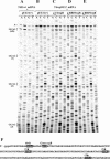
Susceptibility of the T4 transcripts for gene regB to the RegB endoribonuclease of phages T4, RB69 and RB49. Primer extension sequencing of mRNA for gene regB isolated from the E.coli C41(DE3) cells harboring plasmid pET21(+) at 4 min post-infection by T4 wild-type phage at 30°C (A). Primer extension sequencing of the T4regBL52 mRNA for gene regB isolated at 4 min post-infection by phage at 30°C from the E.coli C41(DE3) cells harboring plasmid pET21(+) (B) or the recombinant plasmids pLPT4regB (C), pLTRB69regB (D) and pLPRB49regB (E). Primer Pr. T4regB(R2) was used in the sequencing reactions. The sequencing lanes are labeled with the dideoxynucleotides used in the sequencing reactions. The initiating nucleotides for the transcripts, the GGAG motifs within the SD sequences, as well as the GGAG motifs in the coding region of gene regB mRNA are noted. (F) The nucleotide sequence of the 5′ part of mRNA for gene regB of bacteriophage T4. The GGAG motif within the SD sequence, the initiation codon, as well as the GGAG motifs in the coding region are shown with black backgrounds. Vertical arrows denote the positions of RegB cleavage.

Similar articles

Cited by

References

    1. Ueno H. and Yonesaki,T. (2001) Recognition and specific degradation of bacteriophage T4 mRNAs. Genetics, 158, 7–17. - PMC - PubMed
    1. Miller E.S., Kutter,E., Mosig,G., Arisaka,F., Kunisawa,T. and Rüger,W. (2003) Bacteriophage T4 genome. Microbiol. Mol. Biol. Rev., 67, 86–156. - PMC - PubMed
    1. Otsuka Y., Ueno,H. and Yonesaki,T. (2003) Escherichia coli endoribonucleases involved in cleavage of bacteriophage T4 mRNAs. J. Bacteriol., 185, 983–990. - PMC - PubMed
    1. Deutscher M.P. and Li,Z. (2001) Exoribonucleases and their multiple roles in RNA metabolism. Prog. Nucleic Acid Res. Mol. Biol., 66, 67–105. - PubMed
    1. Zuo Y. and Deutscher,M.P. (2001) Exoribonuclease superfamilies: structural analysis and phylogenetic distribution. Nucleic Acids Res., 29, 1017–1026. - PMC - PubMed

Publication types

MeSH terms

Associated data