

Review
doi: 10.1371/journal.pbio.0020348.
Epub 2004 Oct 12.
Skeletal muscle fiber type: influence on contractile and metabolic properties
Affiliations
- PMID: 15486583
- PMCID: PMC521732
- DOI: 10.1371/journal.pbio.0020348
Item in Clipboard
Review
Skeletal muscle fiber type: influence on contractile and metabolic properties
PLoS Biol.
2004 Oct.
Abstract
Zierath and Hawley discuss how different fiber types affect muscle metabolism and what the signals are that regulate muscle phenotype
Figures
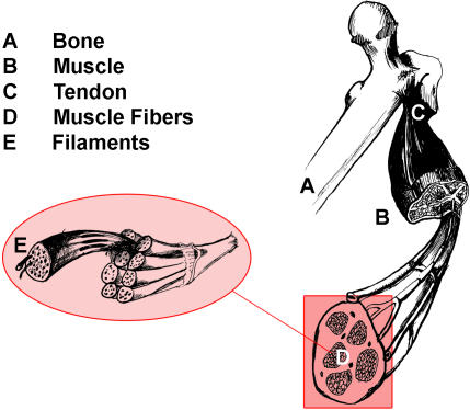
Individual bundles of muscle fibers are called fascicles. The cell membrane surrounding the muscle cell is the sarcolemma, and beneath the sarcolemma lies the sarcoplasm, which contains the cellular proteins, organelles, and myofibrils. The myofibrils are composed of two major types of protein filaments: the thinner actin filament, and the thicker myosin filament. The arrangement of these two protein filaments gives skeletal muscle its striated appearance.
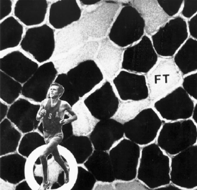
The darkly stained fibers are relatively slow in contractile rate and are ST. These fibers demonstrate a higher aerobic (oxidative) capacity and a lower anaerobic (glycolytic) potential than the lighter stained FT fibers. Shorter's muscle contains approximately 80% ST fibers. Reproduced with kind permission from David L. Costill and William J. Fink.
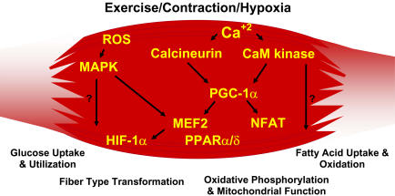
Contraction-induced changes in intracellular calcium or reactive oxygen species provide signals to diverse pathways that include the MAPKs, calcineurin and calcium/calmodulin-dependent protein kinase IV to activate transcription factors that regulate gene expression and enzyme activity in skeletal muscle.
References
-
- Bergstrom J, Hultman E. Muscle glycogen synthesis after exercise: An enhancing factor localized to the muscle cells in man. Nature. 1966;210:309–310. - PubMed
-
- Booth FW, Thomason DB. Molecular and cellular adaptation of muscle in response to exercise: Perspectives of various models. Physiol Rev. 1991;71:541–585. - PubMed
-
- Brooke MH, Kasier KK. Three “myosin ATPase” systems: The nature of their pH liability and sulphydryl dependence. J Histochem Cytochem. 1970;18:670–672. - PubMed
