

Melanoma-restricted genes
- PMID: 15488140
- PMCID: PMC527872
- DOI: 10.1186/1479-5876-2-34
Melanoma-restricted genes
Abstract
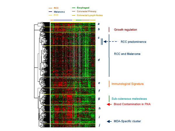
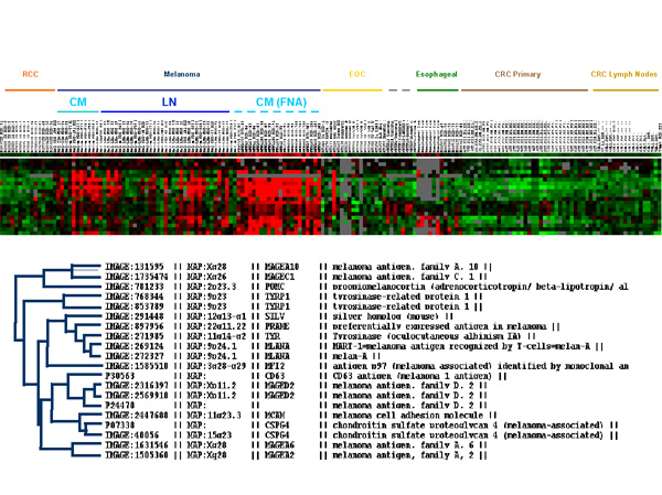
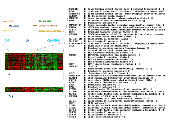
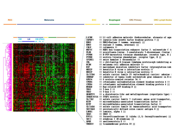
Human metastatic cutaneous melanoma has gained a well deserved reputation for its immune responsiveness. The reason(s) remain(s) unknown. We attempted previously to characterize several variables that may affect the relationship between tumor and host immune cells but, taken one at the time, none yielded a convincing explanation. With explorative purposes, high-throughput technology was applied here to portray transcriptional characteristics unique to metastatic cutaneous melanoma that may or may not be relevant to its immunogenic potential. Several functional signatures could be identified descriptive of immune or other biological functions. In addition, the transcriptional profile of metastatic melanoma was compared with that of primary renal cell cancers (RCC) identifying several genes co-coordinately expressed by the two tumor types. Since RCC is another immune responsive tumor, commonalities between RCC and melanoma may help untangle the enigma of their potential immune responsiveness. This purely descriptive study provides, therefore, a map for the investigation of metastatic melanoma in future clinical trials and at the same time may invite consideration of novel therapeutic targets.
Figures
References
-
- Atkins MB, Lotze MT, Dutcher JP, Fisher RI, Weiss G, Margolin K, Abrams J, Sznol M, Parkinson D, Hawkins M, Paradise C, Kunkel L, Rosenberg SA. High-dose recombinant interleukin-2 therapy for patients with metastatic melanoma: analysis of 270 patients treated between 1985 and 1993. J Clin Oncol. 1998;17:2105–2116. - PubMed
-
- Atkins MB, Sparano J, Fisher RI, Weiss GR, Margolin KA, Fink KI, Rubinstein L, Louie A, Mier JW, Gucalp R. Randomized phase II trial of high-dose interleukin-2 either alone or in combination with interferon alfa-2b in advanced renal cell carcinoma. J Clin Oncol. 1993;11:661–670. - PubMed
-
- Wang E, Marincola FM. cDNA microarrays and the enigma of melanoma immune responsiveness. Cancer J Sci Am. 2001;7:16–23. - PubMed
-
- Kawakami Y, Zakut R, Topalian SL, Stotter H, Rosenberg SA. Shared human melanoma antigens. Recognition by tumor-infiltrating lymphocytes in HLA-A2.1-transfected melanomas. J Immunol. 1992;148:638–643. - PubMed
-
- Kawakami Y, Rosenberg SA. T-cell recognition of self peptides as tumor rejection antigens. Immunol Res. 1996;15:179–190. - PubMed
LinkOut - more resources
Full Text Sources
Other Literature Sources
Miscellaneous
