. 2004 Oct 26;101(43):15452-7.
doi: 10.1073/pnas.0402874101. Epub 2004 Oct 15.

Ceramide, a target for antiretroviral therapy

Affiliations

Ceramide, a target for antiretroviral therapy

Catherine M Finnegan et al. Proc Natl Acad Sci U S A. .

Abstract

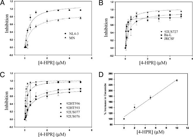
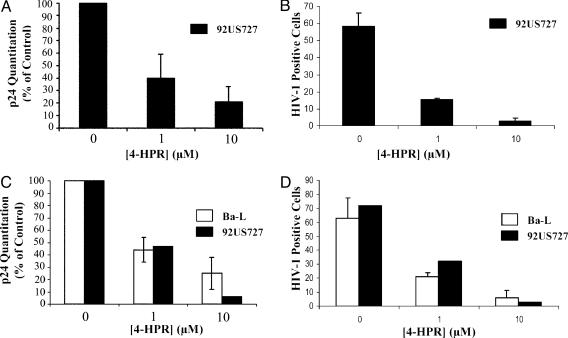
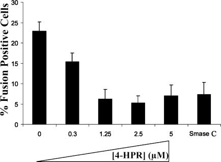
Studies of ceramide metabolism and function in a wide range of biological processes have revealed a role for this lipid in regulating key cellular responses. Our research on the role of sphingolipids in HIV entry has led to the hypothesis that modulation of ceramide levels in target cells affects their susceptibility to HIV infection by rearranging HIV receptors. Cellular ceramide levels were modulated by application of pharmacological agents such as N-(4-hydroxyphenyl)retinamide (4-HPR, fenretinide), by treatment with sphingomyelinase (Smase), or by exogenous addition of long-chain ceramide, and determined after metabolic incorporation of [3H]sphingosine. Infectivity assays were performed by using a HeLa-derived indicator cell line, TZM-bl, CD4+ lymphocytes, and monocytes. We observed a dose-dependent inhibition by 4-HPR of infection of TZM-bl cells by a broad range of HIV-1 isolates at low micromolar concentrations with an IC50 of <1 microM for most isolates tested. Nearly complete inhibition was seen at 5 microM, a dose that enhanced ceramide levels by 50-100%, yet was nontoxic to the cells. Treating cells with other pharmacological agents that enhanced ceramide levels, with Smase, or exogenous addition of long-chain ceramide also resulted in inhibition of HIV-1 infection. Enhancing ceramide levels in CD4+ lymphocytes and in monocyte-derived macrophages with 4-HPR or Smase significantly reduced infectivity without toxicity. The minimal toxicity of normal cells exposed to 4-HPR should make the drug exceedingly suitable as an anti-HIV therapeutic.


Figures

Fig. 1.
Fig. 1.
The use of 4-HPR enhances ceramide levels in TZM-bl cells and blocks HIV-1 infectivity. TZM-bl cells were incubated with 4-HPR for 48 h at 37°C. HIV-1 infection was carried out as described in Experimental Procedures by using the indicated HIV-1 isolate at a multiplicity of infection of 0.01. Luciferase activity was quantified after 16 h, and viral inhibition was calculated relative to control untreated cells. Data are representative of an assay performed in duplicate and are representative of three independent experiments giving similar results. The lines represent best fits to a simple hyperbolic function by using the program sigmaplot. (A) CXCR4 isolates. (B) CCR5 isolates. (C) Dual-tropic primary isolates. (D) Measurement of the increase in TZM-bl ceramide levels after 4-HPR treatment by using the [3H]sphingosine incorporation method described in Experimental Procedures.
Fig. 2.
Fig. 2.
Effect of HPR on HIV-1 infectivity in CD4+ T cells and MDMs. (A and B) Activated CD4+ T cells were treated for 48 h with 4-HPR at the indicated concentration and then infected with HIV-192US727. Infectivity was determined by p24 production (A) and by intracellular viral core staining (B) after 7-12 days as described in Experimental Procedures. Data are representative of three independent experiments. (C and D) Elutriated monocytes were differentiated for 5 days and incubated with 4-HPR for 48 h as described in Experimental Procedures. Infectivity was determined after infection with HIV-1Ba-L or HIV-192US727 by p24 production (C) and by intracellular viral core staining (D). Data are representative of three independent experiments for HIV-1Ba-L infectivity and of one experiment for HIV-192US727 infectivity.
Fig. 3.
Fig. 3.
The use of 4-HPR inhibits viral cell fusion. Target cells were treated with 4-HPR at the indicated concentration for 48 h before the fusion assay. Infection was then carried out for 2 h by using HIV-1 BlaM virions as described in Experimental Procedures. After infection, target cells were loaded with CCF2/AM dye for 1 h, and the BlaM reaction was allowed to proceed for 12 h at room temperature. The percentage of infected cells (number of blue cells per 100 cells) was calculated for 4-HPR-treated untreated target cells and Smase C-treated cells. The results represent an average of two independent experiments. The total number of cells counted for each condition are as follows: Control, 2,208; 0.3 μM 4-HPR, 792; 1.25 μM 4-HPR, 1,857; 2.5 μM 4-HPR, 1,446; 5 μM 4-HPR, 1,540; and Smase, 1,929.
Fig. 4.
Fig. 4.
Chemokine-triggered cell migration. Different concentrations of SDF-1α (Right) or MIP1-β (Left) were placed in the lower wells of the chemotaxis chamber. CD4+ T cells were treated for 2 days with 4-HPR at the indicated concentrations and placed in the upper wells, which were separated from the lower wells by a polycarbonate filter. The results are expressed as chemotaxis index representing the fold increase of migrating cells in response to chemokines over the response to control medium. Significant cell migration (P < 0.05) was detected with 10 ng/ml chemoattractant.

References

    1. Gallo, S. A., Finnegan, C. M., Viard, M., Raviv, Y., Dimitrov, A., Rawat, S. S., Puri, A., Durell, S. & Blumenthal, R. (2003) Biochim. Biophys. Acta 1614, 36-50. - PubMed
    1. Singer, I. I., Scott, S., Kawka, D. W., Chin, J., Daugherty, B. L., DeMartino, J. A., DiSalvo, J., Gould, S. L., Lineberger, J. E., Malkowitz, L., et al. (2001) J. Virol. 75, 3779-3790. - PMC - PubMed
    1. Rawat, S. S., Viard, M., Gallo, S. A., Rein, A., Blumenthal, R. & Puri, A. (2003) Mol. Membr. Biol. 20, 243-254. - PubMed
    1. Hannun, Y. A. & Obeid, L. M. (2002) J. Biol. Chem. 277, 25847-25850. - PubMed
    1. Cremesti, A. E., Goni, F. M. & Kolesnick, R. (2002) FEBS Lett. 531, 47-53. - PubMed

Publication types